
Glial Cell Refresher
- BioSource Faculty
- Dec 11, 2024
- 10 min read
Updated: Jun 23, 2025
Glial cells, once thought to merely provide structural support to neurons, play critical roles in information processing within the nervous system. By supplying neurons with essential raw materials, chemical signals, and structural components, glia significantly influence neuronal function. This post provides an overview of the four main types of glial cells—astrocytes, microglia, oligodendrocytes, and Schwann cells—highlighting their contributions to neural communication, synaptic remodeling, and overall brain health.
Glial cells interact with each other and neurons, playing their role in information processing. By supplying neurons with essential raw materials, chemical signals, and structural components, glia can directly influence the function of neurons (Breedlove & Watson, 2023).

Caption: Astrocytes (violet) play an active role in neuronal communication through synapses and regulation of neural circuit functions, memory, and learning. Graphic © Juan Gaertner/Dreamstime.com.

Click on our podcast icon to listen to this post.

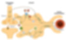
Derived from the Greek word for "glue," the term "glia" reflects a 19th-century assumption that these cells served to "bind the nervous system together." This interpretation has persisted, even though no evidence suggests that glial cells hold nerve cells together. Functions of glial cells that are firmly established include maintaining the ionic environment of nerve cells; influencing the speed of nerve signal propagation; modulating synaptic action via control of neurotransmitter uptake and metabolism at or near the synaptic cleft; providing a framework for certain aspects of neural development; aiding (or in some cases obstructing) recovery from neural injury; forming an interface between the brain and the immune system; and facilitating the convective flow of interstitial fluid through the brain (Purves, 2018). Glial cells significantly differ from neurons despite being at least as numerous. While they don't directly participate in axodendritic and axosomatic synaptic transmission, their support functions are critical in delineating synaptic contacts and maintaining neuronal signaling capabilities. Although many glial cells have intricate processes that extend from their cell bodies, similar to neurons, these are generally less conspicuous and do not fulfill the same roles as neuronal axons and dendrites. Cells exhibiting glial traits seem to be the sole stem cells preserved in the mature brain, capable of generating new glia and, occasionally, new neurons. Although there are hundreds of types of neurons, there are only four main categories of glial cells: astrocytes, microglia, oligodendrocytes, and Schwann cells. Oligodendrocytes constitute over 75% of the brain's glial cells, underscoring myelination's intricate nature and significance. Astrocytes represent approximately 17% of glial cells, while microglia account for a mere 7% of the total (Pelvig et al., 2008). Graphic © Designua/Dreamstime.com

Astrocytes
Astrocytes are star-shaped and are the most prevalent glial cells in the brain. They guide neuronal migration in the embryo and fetus. Since they occupy most of the expanse between neurons and are separated from neurons by nearly 20 µm, they may affect the growth or retraction of axons and dendrites (collectively called neurites). Graphic © Jose Luis Calvo/Shutterstock.com.

An emerging view is that astrocytes help neurons process information and communicate with each other through parallel astrocyte-astrocyte networks. Astrocyte membranes contain neurotransmitter (NT) receptors that can initiate changes in their membrane potential and internal biochemical processes.
Astrocytes directly receive synapses from neurons and integrate neuronal messages, monitor the activity of nearby synapses, communicate with each other using calcium ions and ATP, converse with nearby neurons using NTs, and strongly influence the number of synapses. Schwann cells help determine synapse location.
Astrocytes regulate circulating molecules in the extracellular space (the region surrounding neurons) and the synapse. They enclose synapses, limit the movement of released NTs, and transport them from the synapse to the axon terminal. They also regulate the concentration of ions like potassium outside neurons to prevent interference with their performance (Bear, Connors, & Paradiso, 2020). Graphic © Martin Brož/Dreamstime.com.
Research suggests that interactions between astrocytes and nearby neurons form a tripartite synapse (with "tripartite" denoting a three-part structure), where astrocytes actively participate in the information transfer between neurons (Eroglu & Barres, 2010; Perea et al., 2009). However, the degree and importance of this type of gliotransmission continue to be topics of debate.
Astrocytes transport nutrients, remove wastes, store glycogen during stage-3 sleep, dynamically control local blood flow, and develop and maintain the blood-brain barrier, which protects neurons from toxins. Neurons require astrocytes to access nutrients and oxygen from capillaries. Graphic © Kateryna Kon/Shutterstock.com.

Microglia
Microscopic microglial cells are the primary immune cells of the CNS. They scavenge and engulf diverse materials (phagocytosis), release cytotoxins to control infection, present antigens to T-cells, remove branches from neurons near damaged tissue to aid regrowth, promote tissue repair, and promote chronic neuroinflammation in the CNS that amplifies neurodegeneration. They assist synaptic remodeling by removing unnecessary synapses. Finally, microglia cross the blood-brain barrier to promote homeostasis (Bear, Connors, & Paradiso, 2020). Graphic © Juan Gaertner/Shutterstock.com.

Caption: yellow = neurons, orange = astrocytes, grey = oligodendrocytes, white = microglia.
Neuroprotection
Scheiblich et al. (2024) discovered that microglia use tunneling nanotubules (TNTs) to remove protein aggregates, such as alpha-synuclein and tau, from neurons and to transfer healthy mitochondria to diseased neurons. This process is crucial for maintaining neural function and survival. The study employed RNA sequencing, fluorescent dye tracking, and 3D laser scanning microscopy to observe these interactions in cultured cells and postmortem human brain tissue. Findings indicate that the Rac-PAK pathway is vital for protein aggregate removal via TNTs, and that genetic disruptions associated with neurodegenerative diseases hinder this process. This research sheds light on potential therapeutic targets for neurodegenerative diseases by leveraging the neuroprotective functions of microglia. TNT graphic is reproduced from Microglia rescue neurons from aggregate-induced neuronal dysfunction and death through tunneling nanotubes under the Creative Commons license.

COVID-19
A postmortem study by Schwabenland et al. (2021) provided evidence that COVID-19 may damage the nervous system through CD-8 and CD-4 T-cell-microglial interactions that breach the blood-brain barrier and damage the brain stem and olfactory bulb axons.
Chronic Pain
Following peripheral nerve damage (such as a back injury or amputated limb), microglial cells encircle the synapses between pain fibers and neurons in the spinal cord's dorsal horn. These microglial cells release substances like cytokines, chemokines, ATP, and brain-derived neurotrophic factor (BDNF) that render the dorsal horn neurons hyperresponsive (Coull et al., 2005; Inoue & Tsuda, 2018; Scholz & Woolf, 2007).
As a result, these neurons become persistently active, inundating the thalamus with action potentials that signal pain. Additional immune responses, including inflammation, are also suspected to contribute to neuropathic pain (Ji et al., 2016).
Oligodendrocytes
Oligodendrocytes, which are smaller than astrocytes, form up to 50 segments of myelin that only insulate adjacent axons within the brain and spinal cord of the central nervous system. Myelination continues in selected brain regions for 10-15 years following birth, possibly into adulthood (Breedlove & Watson, 2023).
Myelination Advantages
Myelination involves the wrapping of myelin, a fatty insulating layer, around the axons of nerve cells by oligodendrocytes and Schwann cells. This provides several significant advantages:
Rapid Signal Transmission: By insulating the axon, myelin reduces electrical resistance and increases the speed at which action potentials, or nerve impulses, travel along the neuron. Compare action potential transmission in unmyelinated (top) and myelinated (bottom) axons. The animation © viktorov.pro/Shutterstock.com.
Saltatory Conduction: Myelination enables saltatory conduction, where the action potential jumps between gaps in the myelin called nodes of Ranvier. This further enhances the velocity of signal transmission.
Energy Efficiency: Myelinated neurons require less energy to transmit signals. The ion channels are concentrated at the nodes of Ranvier, limiting the places where ions are exchanged. This means the cell expends less energy maintaining the ion gradients essential for signal transmission.
Protection and Stability: The myelin sheath also adds physical protection to the axons, safeguarding them from mechanical damage and potentially contributing to the overall stability of the neural network.
Enhanced Communication Precision: By preventing the leakage of electrical signals, myelination helps maintain the fidelity of the signal, allowing for more precise communication between neurons.
Facilitation of Complex Functions: The increased efficiency and speed of communication between neurons through myelination enable more complex neurological functions, contributing to higher cognitive processes in humans.
Overall, myelination plays a crucial role in optimizing the function, speed, and energy efficiency of neurons, greatly enhancing the complexity and functionality of the nervous system.
Oligodendrocytes release trophic factors that promote axonal structural integrity (Nave, 2010). They block axonal regeneration by releasing growth-inhibitory proteins following injury. These molecules are part of the reason for minimal functional recovery in the CNS following spinal cord damage.
Multiple sclerosis, a demyelinating disease, destroys oligodendrocytes.
Schwann Cells
Schwann cells provide myelin for single PNS axons and facilitate axonal regeneration following damage (Breedlove & Watson, 2023). Graphic © Designua/Shutterstock.com.

Conclusion
Glial cells are integral to the nervous system's function, going beyond structural support to actively participate in neural communication, synaptic remodeling, and overall brain health. Astrocytes, microglia, oligodendrocytes, and Schwann cells each play unique roles in maintaining and enhancing neuronal function. Understanding these roles opens new avenues for therapeutic interventions in neurodegenerative diseases and neural injuries, leveraging the diverse capabilities of glial cells to support and protect neurons.
Google Illuminate Discussion
Please click on the podcast icon below for an engaging Google Illuminate discussion.

Glossary
adenosine triphosphate (ATP): a molecule that carries energy within cells. It's essential for all the body's processes that require energy.
alpha-synuclein: a protein predominantly found in the brain, where it plays a role in synaptic function and neurotransmitter release. In neurodegenerative diseases such as Parkinson's disease, alpha-synuclein can misfold and aggregate, forming toxic clumps that contribute to neuronal damage and death. The accumulation of alpha-synuclein aggregates is a hallmark of several neurodegenerative conditions, and their clearance is vital for neuronal health.
astrocytes: star-shaped glial cells in the brain and spinal cord, involved in many functions such as supporting neuronal functioning, regulating neurotransmission, and maintaining the blood-brain barrier.
blood-brain barrier: a semipermeable border that separates the circulating blood from the brain and extracellular fluid in the central nervous system.
brain-derived neurotrophic factor (BDNF): a protein that promotes survival, growth, and the formation of new synapses by neurons, which may remodel the dorsal horn in neuropathic pain.
capillaries: the smallest blood vessels in the body that enable the exchange of water, oxygen, carbon dioxide, and nutrients between blood and tissues.
chemokines: small proteins that induce directed chemotaxis (migration) in nearby responsive cells, playing roles in immune responses and inflammation.
cytokines: proteins that are important in cell signaling, affecting the behavior of cells around them.
glial cells: non-neuronal cells that maintain homeostasis, form myelin, and provide support and protection for neurons in the central and peripheral nervous system. Unlike neurons, glial cells do not carry electrical impulses. There are several types of glial cells, including astrocytes, oligodendrocytes, microglia, and Schwann cells, each with unique functions such as regulating neurotransmission, mounting immune responses, forming myelin, and aiding in neural development and repair.
gliotransmission: the process by which glial cells communicate with neurons, contributing to synaptic transmission.
microglia: a glial cell that acts as the main form of active immune defense in the central nervous system. multiple sclerosis (MS): a chronic disease primarily involving the central nervous system where the immune system mistakenly attacks the protective covering of nerve fibers (myelin), causing communication problems between the brain and the rest of the body. This can lead to permanent damage or deterioration of the nerves. Symptoms vary widely and may include fatigue, difficulty walking, numbness or tingling, muscle weakness, and problems with coordination and balance.
neuropathic pain: pain caused by damage or disease affecting the somatosensory nervous system.
node of Ranvier: a gap in the myelin sheath of a nerve between adjacent Schwann cells.
oligodendrocytes are central nervous system glial cells forming a myelin sheath around axons.
peripheral nervous system: the PNS consists of all nerves and neurons outside the brain and spinal cord. This network of nerves extends to the limbs and organs and connects the CNS to the rest of the body, enabling it to respond appropriately to stimuli. The PNS is further subdivided into the somatic nervous system, which controls voluntary muscle movement and processes sensory information, and the autonomic nervous system, which controls heart rate, digestion, and respiration rate.
Rac-PAK pathway: a signaling cascade involved in the regulation of cytoskeletal dynamics, cell migration, and various cellular responses to external stimuli. Rac is a small GTPase that, when activated, interacts with PAK (p21-activated kinase) to modulate actin cytoskeleton reorganization, which is essential for processes such as cell motility and the formation of cellular structures like TNTs. This pathway is critical for the removal of protein aggregates from neurons via TNTs, highlighting its importance in neuroprotective mechanisms.
Schwann cells: peripheral nervous system glial cells that produce the myelin sheath around neuronal axons.
synaptic remodeling: the process by which synapses are strengthened, weakened, created, or removed in response to neuronal activity.
tau: a protein associated with microtubules in neurons, where it stabilizes these structures and supports their function in intracellular transport. In conditions like Alzheimer's disease, tau becomes hyperphosphorylated and forms insoluble aggregates known as neurofibrillary tangles. These tau aggregates disrupt neuronal function and contribute to cell death. Efficient removal of tau aggregates is essential for preventing and mitigating neurodegenerative processes.
tripartite synapse: a concept that expands the traditional neuron-neuron synaptic cleft to include interactions with glial cells, specifically astrocytes.
tunneling nanotubes (TNTs): thin, tubular structures that form connections between cells, allowing for direct cell-to-cell communication and the transfer of various cellular components, such as proteins, organelles, and other molecules. In the context of neurobiology, TNTs enable the exchange of materials between microglia and neurons, playing a crucial role in maintaining neuronal health and function, particularly by facilitating the removal of toxic protein aggregates and the delivery of healthy mitochondria.
References
Coull, J. A., Beggs, S., Boudreau, D., Boivin, D., Tsuda, M., Inoue, K., Gravel, C., Salter, M. W., & De Koninck, Y. (2005). BDNF from microglia causes the shift in neuronal anion gradient underlying neuropathic pain. Nature, 438(7070), 1017–1021. https://doi.org/10.1038/nature04223 Eroglu, C., and Barres, B. A. (2010). Regulation of synaptic connectivity by glia. Nature, 468(7321), 223–231. Inoue, K., & Tsuda, M. (2018). Microglia in neuropathic pain: Cellular and molecular mechanisms and therapeutic potential. Nat. Rev. Neurosci., 19(3), 138–152.
Ji, R. R., Chamessian, A., and Zhang, Y. Q. (2016). Pain regulation by non-neuronal cells and inflammation. Science, 354(6312), 572–577.
Nave, K. A. (2010). Myelination and the trophic support of long axons. Nat. Rev. Neurosci., 11, 275–283.
Pelvig, D. P., Pakkenberg, H., Stark, A. K., and Pakkenberg, B. (2008). Neocortical glial cell numbers in human brains. Neurobiol. Aging, 29, 1754–1762.
Perea, G., Navarrete, M., and Araque, A. (2009). Tripartite synapses: Astrocytes process and control synaptic information. Trends Neurosci., 32, 421–431.
Scholz, J., & Woolf, C. J. (2007). The neuropathic pain triad: Neurons, immune cells and glia. Nature Neuroscience, 10(11), 1361-1368. Scheiblich, H., Eikens, F., Wischhof, L., Opitz, S., Jüngling, K., Cserép, C., Schmidt, S. V., Lambertz, J., Bellande, T., Pósfai, B., Geck, C., Spitzer, J., Odainic, A., Castro-Gomez, S., Schwartz, S., Boussaad, I., Krüger, R., Glaab, E., Di Monte, D. A., Bano, D., … Heneka, M. T. (2024). Microglia rescue neurons from aggregate-induced neuronal dysfunction and death through tunneling nanotubes. Neuron, S0896-6273(24)00491-4. Advance online publication. https://doi.org/10.1016/j.neuron.2024.06.029 Schwabenland, M., Salié, H., Tanevski, J., Killmer, S., Lago, M. S., Schlaak, A. E., Mayer, L., Matschke, J., Püschel, K., Fitzek, A., Ondruschka, B., Mei, H. E., Boettler, T., Neumann-Haefelin, C., Hofmann, M., Breithaupt, A., Genc, N., Stadelmann, C., Saez-Rodriguez, J., Bronsert, P., … Bengsch, B. (2021). Deep spatial profiling of human COVID-19 brains reveals neuroinflammation with distinct microanatomical microglia-T-cell interactions. Immunity, 54(7), 1594–1610.e11. https://doi.org/10.1016/j.immuni.2021.06.002
Please Support Our Friends


