
5-Second Science: Connectome 2.0 Achieves Live Near-Microscopic Brain Imaging
- Fred Shaffer
- Sep 1, 2025
- 4 min read


Connectome 2.0
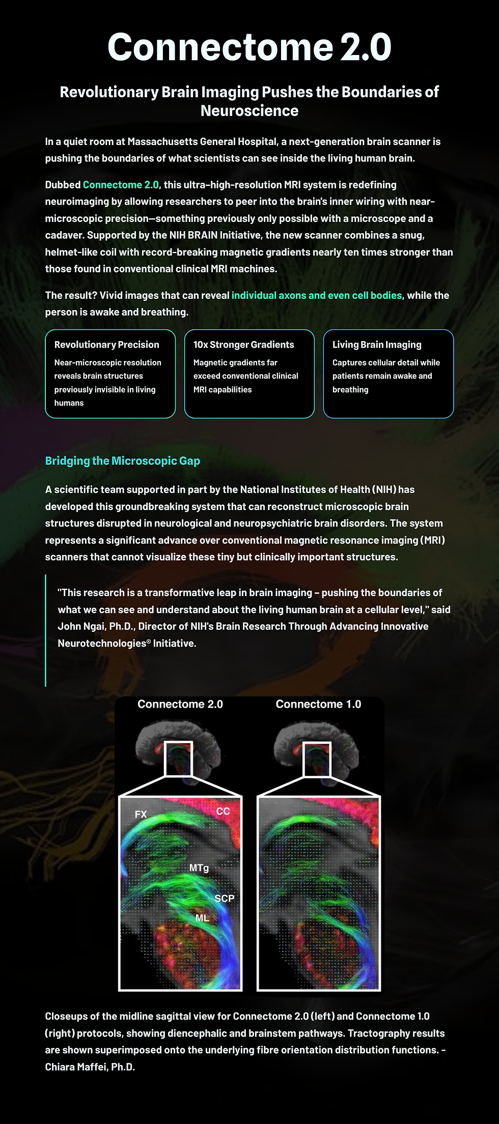
In a quiet room at Massachusetts General Hospital, a next-generation brain scanner is pushing the boundaries of what scientists can see inside the living human brain.

Dubbed Connectome 2.0, this ultra–high-resolution MRI system is redefining neuroimaging by allowing researchers to peer into the brain’s inner wiring with near-microscopic precision—something previously only possible with a microscope and a cadaver. Supported by the NIH BRAIN Initiative, the new scanner combines a snug, helmet-like coil with record-breaking magnetic gradients nearly ten times stronger than those found in conventional clinical MRI machines. The result? Vivid images that can reveal individual axons and even cell bodies, while the person is awake and breathing.

A scientific team supported in part by the National Institutes of Health (NIH) has developed a new, ultra-high-resolution brain imaging system that can reconstruct microscopic brain structures that are disrupted in neurological and neuropsychiatric brain disorders. The new system is a significant advance over conventional magnetic resonance imaging (MRI) scanners that cannot visualize these tiny but clinically important structures.
The system, called the Connectome 2.0 human MRI scanner, overcomes a significant hurdle for neuroscientists: being able to bridge different brain regions and probe tiny structures necessary to define the “connectome,” the complex matrix of structural connections between nodes in the nervous system, and to do it noninvasively in living humans.
“This research is a transformative leap in brain imaging – pushing the boundaries of what we can see and understand about the living human brain at a cellular level,” said John Ngai, Ph.D., Director of NIH’s Brain Research Through Advancing Innovative Neurotechnologies Initiative®, or The BRAIN Initiative®. “The new scanner lays essential groundwork for the BRAIN CONNECTS program’s ultimate goal of developing a wiring diagram for the human brain.”
The scanner is innovative in two major ways: it fits snugly around the heads of living people, and it has many more channels than
But the Connectome 2.0 is more than just a sharper camera—it’s a tool for decoding the brain’s deepest secrets. Early volunteer scans have already begun, and researchers are distributing access to the system through the BRAIN CONNECTS U24 program, offering outside scientists a rare opportunity to explore the living human connectome in stunning detail. Ultimately, this innovation could speed up the search for answers in Alzheimer’s, autism, multiple sclerosis, and other disorders where the brain’s architecture goes awry. For the first time, we may be able to watch these changes unfold at the cellular level in real time, ushering in a new era of precision neuroscience.
Thanks to Michael Sitar for sharing this NIH announcement.
Key Takeaways
Connectome 2.0 is an advanced MRI scanner that visualizes microscopic brain structures in living humans with near-cellular resolution.
The system uses ultra-strong magnetic gradients (up to 500 mT/m) and a high-density head coil to achieve unprecedented signal clarity and spatial detail.
It enables noninvasive imaging of axons and cell bodies, previously only visible in postmortem or animal studies.
The technology supports the NIH BRAIN Initiative, offering researchers access through the BRAIN CONNECTS U24 program.
Connectome 2.0 may transform neurological research, enabling real-time study of disorders like Alzheimer’s, autism, and multiple sclerosis.

Glossary
axon: the long, slender projection of a neuron that conducts electrical impulses away from the neuron's cell body.
connectome: the complete map of neural connections within the brain, often represented as a network of nodes (regions) and edges (pathways).
diffusion MRI: a type of MRI that tracks the diffusion of water molecules, used to infer microstructural features like fiber orientation and axon diameter.
field monitoring: a technique that measures temporal and spatial variations in the MRI’s magnetic field to correct distortions in the resulting images.
gradient coil: components in an MRI that produce controlled, varying magnetic fields (gradients); stronger gradients improve spatial resolution and diffusion sensitivity.
micron: a unit of length equal to one millionth of a meter (1 µm), used to specify the high resolution achieved by the Connectome 2.0 scanner.
peripheral nerve stimulation (PNS): unwanted stimulation of peripheral nerves caused by rapidly switched magnetic fields in strong-gradient MRI systems.
RF coil (radiofrequency coil): the antenna used to send and receive radiofrequency signals in MRI; higher channel count improves signal reception and image clarity.
signal-to-noise ratio (SNR): a measure of signal strength relative to background noise; higher SNR yields clearer and more detailed imaging.
Reference
https://www.nih.gov/news-events/scientists-develop-high-performance-mri-scanner-effort-define-microscopic-brain-structures
About the Author

Fred Shaffer earned his PhD in Psychology from Oklahoma State University. He earned BCIA certifications in Biofeedback and HRV Biofeedback. Fred is an Allen Fellow and Professor of Psychology at Truman State University, where has has taught for 50 years. He is a Biological Psychologist who consults and lectures in heart rate variability biofeedback, Physiological Psychology, and Psychopharmacology. Fred helped to edit Evidence-Based Practice in Biofeedback and Neurofeedback (3rd and 4th eds.) and helped to maintain BCIA's certification programs.
Support Our Friends


Comments