
Not a Fear Center: How the Networked Amygdala Changes Neurofeedback Practice

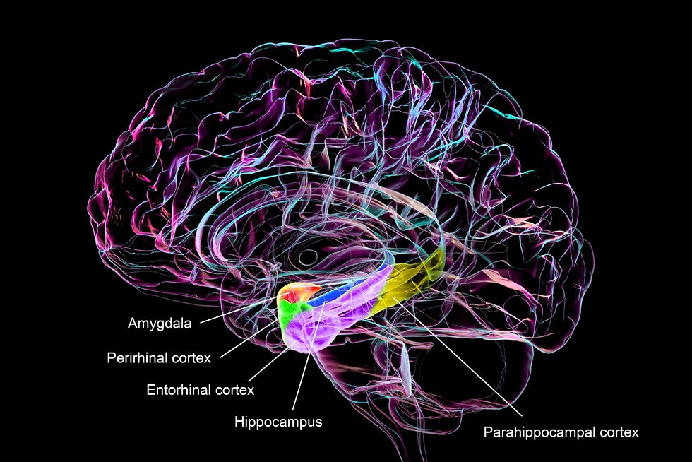
Many clients arrive believing that an overactive amygdala explains their anxiety, and some neurofeedback protocols rest on the same belief. In one face-matching study, every subregion of the amygdala, a cluster of cell groups deep in each temporal lobe, responded to happy faces as strongly as to fearful ones (Labuschagne et al., 2024). Its subdivisions join separate large-scale brain networks (Elvira et al., 2022).

Fox and Shackman (2024) concluded in a review of human and animal research that the amygdala should not be treated as a fear and anxiety center. We draw only on human studies to show what the evidence supports in place of the fear-center model and what that shift means for clinicians who train the brain.
Retire the Fear Center: Human Data Show Several Context-Dependent Roles
The older model treated the amygdala as a single alarm that fires in proportion to fear. Labuschagne et al. (2024) scanned 86 healthy adults during a single face-matching task with fearful, angry, and happy faces. Every amygdala subregion they examined responded to all three expressions, with no difference between emotions. The fear-specific signal appeared instead in connectivity, as stronger coupling between the basolateral amygdala (BLA) and face-processing cortex for fearful faces. The authors concluded that fear specialization may not show up in local amygdala activity at all.
Wang et al. (2017) recorded 234 single amygdala neurons in nine neurosurgical patients viewing faces morphed between fear and happiness. About 14% of the neurons tracked expression intensity, and a similar share tracked ambiguity. Three patients with bilateral amygdala lesions judged faces as fearful at a lower threshold than controls did.
Jang and Kragel (2025) trained artificial neural network models on functional magnetic resonance imaging (fMRI) data from 20 adults watching one film, then tested them on standardized image sets. The models tracked valence, how pleasant or unpleasant an image felt, but not arousal, its emotional intensity. The authors proposed that the amygdala compresses rich sensory input into a few behaviorally relevant dimensions, an interpretation that later studies must test. No single replacement slogan fits these findings. Human evidence supports several context-dependent functions involving different nuclei and broader networks.
Each Amygdala Subdivision Works With Its Own Network Partners
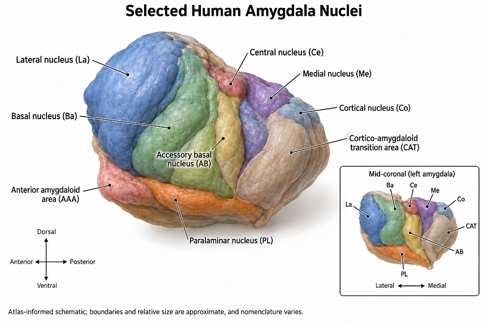
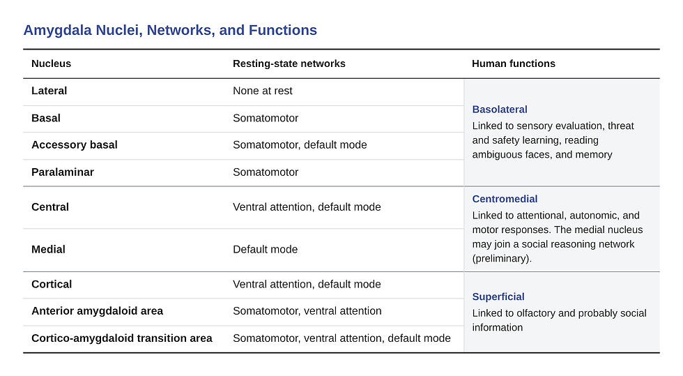
A nucleus is a cluster of nerve cells that share connections. For clinical purposes, three broad amygdala groups matter: the BLA, the centromedial amygdala (CMA), and the superficial amygdala. Bzdok et al. (2013) used thousands of published imaging experiments to map which tasks co-activate each group. The BLA was associated with high-level sensory input, the CMA with attentional, autonomic, and motor responses, and the superficial group with olfactory and probably social information.
Direct stimulation in patients fits that broad division. Zhang et al. (2023) stimulated 152 amygdala contacts in 48 patients with drug-resistant epilepsy and recorded 250 responses. Half were autonomic responses, and they arose from almost every subdivision. Basolateral sites mainly evoked emotional feelings, bodily sensations, and vestibular sensations, while superficial sites evoked emotions and olfactory or visual hallucinations.
Elvira et al. (2022) analyzed 7-tesla resting scans from 172 adults to see which nuclei move with which networks. They found three configurations linked to the somatomotor network, the ventral attention network, and the default mode network. Each network drew on a different combination of nuclei, and the lateral nucleus did not join any of them. Matyi et al. (2021) analyzed white matter connections in 1,052 adults and concluded that there is no single "amygdala network."
Edmonds et al. (2024) mapped functional connectivity, the correlation of slow signal fluctuations between regions, within each of six adults on a 7-tesla scanner.

The network people use when reasoning about others' minds included regions near the BLA and the medial nucleus in most participants. The regions did not appear in 3-tesla data, so this social network finding remains preliminary.
The network view gives clinicians a better map than the alarm metaphor. Autonomic responses, attention to salient events, internally focused thought, and social reasoning draw on different amygdala partnerships (Edmonds et al., 2024; Elvira et al., 2022; Zhang et al., 2023).
These are group-level associations, so they cannot show which network drives a particular client's symptoms. Treat a symptom pattern as a prompt for assessment and a working hypothesis to test, and keep the anatomy at three groups and several networks.
Amygdala Threat Responses Depend on Timing and Task Design
Pavlovian threat conditioning pairs a neutral cue with an aversive event until the cue alone triggers a defensive response. Wen et al. (2022) pooled fMRI data from 601 participants, including 114 with posttraumatic stress disorder (PTSD) and 92 with anxiety disorders. Averaged over all trials, the amygdala response to the threat cue was negligible. Over the first four trials, it was about four times larger, a moderate effect, and it then faded rapidly.
Both the BLA and the CMA responded early, and the CMA coupled with the insula and the dorsal anterior cingulate cortex (dACC). Late in learning, the BLA reversed direction and responded more to the safe cue, coupling with the hippocampus and the ventromedial prefrontal cortex (vmPFC). The authors read that reversal as a safety signal.
Radua et al. (2025) ran a meta-analysis that pooled participant-level data across studies from 2,199 participants in 21 laboratories. Among 1,888 healthy participants, neither amygdala showed a reliable average difference between threat and safety cues, and both differences were tiny.
The right amygdala responded earlier than the left in learning, while the insula and the dACC responded reliably. Visser et al. (2021) pooled three studies with 98 participants and found strong amygdala responses to faces but none to the threat cue. Signal dropout was low, so the authors rejected poor signal as the explanation.
Wen et al. (2024) used multivariate pattern analysis, which tests whether activity patterns across many small imaging units can distinguish two conditions. In 1,465 participants, whole-brain patterns that excluded the classic threat circuit decoded threat versus safety cues with 77.5% to 88.6% accuracy across conditioning blocks. The classic circuit alone reached 65.5% to 74.0%, though the authors reported no test of that difference.
Taken together, amygdala findings in threat learning vary with trial timing and task design rather than following one universal time course (Radua et al., 2025; Visser et al., 2021; Wen et al., 2022).
Meijer et al. (2026) applied transcranial ultrasound stimulation (TUS), which focuses low-intensity sound waves through the skull, to the BLA of 25 healthy adults. Compared with sham cues in the same session, amygdala TUS moderately slowed early threat learning, measured by the skin conductance response (SCR). It sped early extinction learning, the new learning that occurs when a cue stops predicting harm, by a similar amount. A separate group receiving TUS to the hippocampus showed no specific effects. These results point to an amygdala role in early learning within one conditioning task, and other threat situations may differ.
Some People Panic Without a Working Amygdala
Bechara et al. (1995) found that a patient with bilateral amygdala damage did not acquire conditioned autonomic responses, yet could state which cues predicted the aversive event. A patient with hippocampal damage, a structure needed for consciously reportable memory, showed the reverse pattern.
Adolphs et al. (1995) found that patient SM, who has bilateral amygdala damage, rated fearful faces far less intense than controls did, whereas patients with one-sided damage did not.
Feinstein et al. (2011) took SM through a pet store with live snakes, a haunted house, and 10 horror film clips. Her fear ratings never exceeded 2 on a 0-10 scale, and 3 months of experience sampling showed almost no fear in daily life.
Anderson and Phelps (2002) asked 20 patients with one-sided amygdala lesions and one patient with bilateral lesions to rate their everyday moods. Their positive and negative moods matched those of controls in both size and frequency.
Feinstein et al. (2013) gave SM and two other women with bilateral amygdala damage from Urbach-Wiethe disease, a rare genetic disorder, one breath of 35% carbon dioxide. All three had panic attacks, compared with 3 of 12 healthy comparison women.
Khalsa et al. (2016) then raised heart rate in two of these women with isoproterenol, a drug that mimics adrenaline's effects on the heart. Both became anxious, and one had a full panic attack, compared with 4 of 15 controls. The authors concluded that the amygdala is not required for panic triggered from within the body and proposed contributions from the brainstem and hypothalamus.
These few cases of experimentally induced panic show only that an intact amygdala is not necessary for every panic response; how often body-driven panic engages it remains open. Assess breathing, heart sensations, and interoception, the sense of the body's internal state, directly rather than treating panic as an amygdala alarm.
The Amygdala Also Reads Ambiguity, Weighs Rewards, and Tracks Other People
Algermissen et al. (2026) applied TUS to the BLA of healthy volunteers before they chose to approach or avoid happy, neutral, and angry faces. Afterward, participants approached neutral faces more often, treating these ambiguous expressions more like happy ones. Sun et al. (2023) found that amygdala neurons coding facial ambiguity responded at about 658 ms, before dorsomedial prefrontal neurons at about 893 ms.
The amygdala also tracks outcomes and other people. Manssuer et al. (2024) recorded reward and punishment signals from amygdala electrodes in 17 patients during a rule-switching task. Aquino et al. (2020) found separate amygdala neurons encoding expected value for oneself and for another person.
In clients, amygdala-related difficulties could plausibly show up as misreading neutral faces, poor learning from feedback, or trouble learning from others, though no clinical study has tested that link. Ask about these patterns during intake as part of a broad assessment.
Emotional Memory: Amygdala Stimulation Nudges What Lasts Without Producing Feelings
Qasim et al. (2023) recorded intracranial activity in 148 patients and found that amygdala and hippocampal activity during study predicted later recall of high-arousal words. Direct electrical stimulation delivers brief current pulses through electrodes implanted for clinical reasons. Inman et al. (2018) stimulated the BLA for 1 s after half of 160 object images in 14 patients, and next-day recognition of the stimulated objects improved substantially. No patient reported feeling the stimulation or any emotion.
Hollearn et al. (2025) pooled 31 patients, including the original 14, and found a much smaller benefit, about a quarter the size of the original estimate. Some patients' memory got worse. Wahlstrom et al. (2026) found a benefit for objects but not scenes, though a direct comparison fell short of statistical significance, so chance remains possible. Amygdala stimulation can nudge which memories last without producing a feeling, but its size and direction vary across people.
A Brain Scan Will Not Diagnose Your Client
Test-retest reliability asks whether a measure gives the same answer when the same person is tested twice. Elliott et al. (2020) combined 90 substudies and found that common task-fMRI measures had poor reliability on average. Nord et al. (2017) scanned 29 healthy volunteers twice and found strong group-level amygdala activation, yet individual amygdala measures ranged from no consistency at all to only modest consistency across sessions.
Tamm et al. (2022) found no association between amygdala responses to negative faces and depressive symptoms in 28,638 UK Biobank adults. Knaust et al. (2025) found no differences in amygdala nucleus volumes among 185 military personnel diagnosed with PTSD, major depressive disorder (MDD), both, or adjustment disorder. That retrospective study had no healthy control group.
Fox and Shackman (2024) reviewed prospective human studies and found that amygdala reactivity predicted later symptoms only weakly, accounting for roughly 3% of the differences between people. They judged that effect too weak for screening, diagnosis, or other clinical use. Assess the person, not the structure. Record the trigger, the client's expectation, the physiological response, its time course, and its cost to daily functioning, and track those across sessions.
What Amygdala Neurofeedback Has Delivered So Far
Real-time fMRI neurofeedback (rtfMRI-NF) displays a person's own signal from a target region within seconds, so the person can learn to change it. Young et al. (2017) randomized 36 unmedicated adults with MDD to two sessions of left amygdala upregulation during positive memories or feedback from a parietal control region. Scores on the Montgomery-Åsberg Depression Rating Scale (MADRS) fell from 23.5 to 11.9 in the amygdala group and from 23.8 to 21.9 in the control group. Follow-up lasted only 1 week.
Later trials have been less encouraging. Zotev et al. (2018) trained combat veterans with PTSD to raise amygdala activity, and the difference from sham in change on the Clinician-Administered PTSD Scale (CAPS) was not significant. Zhao et al. (2023) ran a preregistered, double-blind trial of amygdala downregulation with 14 active and 11 sham completers. CAPS-5 scores improved in both arms, with no group difference at 30 or 60 days. Misaki et al. (2025) pooled 95 adults with MDD and found that the advantage over the control condition on the MADRS was not statistically reliable.
The amygdala electrical fingerprint (EFP) is a signal model that estimates amygdala activity from electroencephalography (EEG), which records voltage at the scalp (Keynan et al., 2019). Keynan et al. randomized 180 healthy male soldiers to six EFP sessions, control EEG feedback, or no feedback. EFP training reduced alexithymia, difficulty identifying and describing one's feelings, more than control feedback did, though the advantage was small and rested on a lenient statistical test. State anxiety did not differ between groups.
Fruchtman-Steinbok et al. (2021) randomized 59 patients with PTSD to EFP training guided by a trauma script, EFP training with a neutral script, or no feedback. The trauma-guided group improved more on the CAPS-5 than the neutral-script group, but the difference fell short of statistical significance. Fine et al. (2024) added EFP sessions to therapy for 55 women with treatment-resistant PTSD, and the primary comparison narrowly missed statistical significance. In both trials, how well patients learned to regulate the EFP signal did not correlate with symptom change (Fine et al., 2024; Fruchtman-Steinbok et al., 2021).
Fruchter et al. (2024) ran a single-arm, open-label trial of EFP neurofeedback at five sites in 79 adults with chronic PTSD. Among 66 participants in the effectiveness analysis, 66.7% showed a clinically meaningful CAPS-5 reduction at 3 months. Without a control group, the trial cannot show that neurofeedback caused this improvement, as the authors acknowledged. Several authors reported financial ties to the company that makes the EFP software.
Berman et al. (2025) meta-analyzed nine randomized trials of neurofeedback for PTSD. Two fMRI trials against sham showed essentially no average advantage, but the plausible results ranged from a moderate benefit to a moderate disadvantage, leaving substantial uncertainty. The larger EEG effects came from comparisons with passive controls and carried very low certainty.
Goldway et al. (2022) found that people can learn to modulate amygdala signals, which makes clinical transfer a plausible bottleneck. Where amygdala neurofeedback is available, offer it as an experimental adjunct with informed consent, and judge it by measured symptoms.
Training Pathways: A Promising Hypothesis for Neurofeedback
If symptoms involve network partnerships, a single-region target may be the wrong unit, and early trials have begun to test that idea. Young et al. (2018) reanalyzed the 2017 depression trial and found that amygdala training increased amygdala connectivity with medial prefrontal, insular, cingulate, and striatal regions. Changes in several of these connections explained significant variance in symptom improvement. Misaki et al. (2025) likewise found that clinical response tracked whole-brain activation patterns during training rather than left amygdala activation.
Connectivity-based neurofeedback rewards coupling between two regions rather than activity in one. Koush et al. (2017) used dynamic causal modeling, which estimates the direction of influence between regions, to give nine healthy adults feedback on top-down prefrontal influence over the amygdala. They learned to strengthen it, while six sham participants did not, and their ratings of emotional pictures became more positive. Questionnaire measures of anxiety and depression did not change, and the groups were very small.
In a randomized crossover study, Zhao et al. (2019) gave 26 healthy adults with high anxiety real and sham feedback on coupling between the amygdala and the ventrolateral prefrontal cortex. Real training strengthened that pathway and reduced anxiety ratings, whereas sham training did neither. Anxiety did not change at a follow-up session 3 days later. Keynan et al. (2019) also reported greater amygdala–vmPFC connectivity after EFP training than after no training.
Heart rate variability (HRV) describes beat-to-beat change in heart rate and reflects autonomic modulation. Thayer et al. (2012) meta-analyzed neuroimaging studies and found that HRV was associated with regional cerebral blood flow in areas including the amygdala and the vmPFC. Sakaki et al. (2016) measured RMSSD, the root mean square of successive differences between heartbeats. Higher RMSSD was associated with stronger resting connectivity between the right amygdala and the medial prefrontal cortex (mPFC).
HRV biofeedback teaches slow, paced breathing that increases heart rate oscillations. Nashiro et al. (2023) randomized 106 healthy young adults to 5 weeks of practice that increased (Osc+) or decreased (Osc−) those oscillations. The preregistered primary outcome, right amygdala–mPFC connectivity, did not differ between conditions, but left amygdala–mPFC connectivity increased in Osc+. Cho et al. (2023) reported that this connectivity change statistically mediated a more positive memory bias, an indirect-effect analysis that does not establish causation on its own. Measures of anxiety, depression, and mood did not differ between conditions (Nashiro et al., 2023).
Implanted devices push the network logic further. Gill et al. (2023) reported a pilot in which a device stimulated the amygdala only when it detected sustained 5 to 9 Hz theta activity. CAPS-5 scores improved 87.65% and 36.84% in two men with treatment-resistant PTSD. Koek et al. (2024) reported 44% and 55% improvement in two veterans after 4 years of continuous BLA deep brain stimulation (DBS). With four patients across two reports, these results show feasibility only.
These studies point toward pathway targets, but the evidence is early. Connectivity training has worked mainly in small healthy samples, and its symptom effects have been brief or absent (Koush et al., 2017; Zhao et al., 2019). Matching a target to a client's presumed network problem, such as prefrontal regulation of the amygdala for emotion regulation, remains a research hypothesis.
None of the trials reviewed here show that pathway-based training produces better clinical outcomes than single-region training. Measure symptoms and daily functioning at baseline and after training, and treat any network change as skill acquisition rather than as proof of clinical benefit.
Integrative Summary: From Alarm to Network Hub
The amygdala is no longer best described as a fear center, and no single slogan has replaced that label. It responds to happy as well as threatening faces, codes intensity and ambiguity, carries reward and social-value signals, and modulates memory without producing feelings (Aquino et al., 2020; Inman et al., 2018; Labuschagne et al., 2024; Manssuer et al., 2024; Wang et al., 2017).
Its subdivisions participate in different networks, from autonomic and attentional systems to default mode and social networks (Elvira et al., 2022; Zhang et al., 2023). Its threat response depends on trial timing and task design, and experimentally induced panic has occurred in a few people without it (Khalsa et al., 2016; Radua et al., 2025; Wen et al., 2022).
For neurofeedback, the lesson is to train what the evidence supports and to measure what matters to clients. Amygdala signals are learnable but unreliable as individual diagnostics, and single-region amygdala training has not consistently beaten sham on symptoms (Berman et al., 2025; Nord et al., 2017). Connectivity targets fit the network model, but whether they improve clinical outcomes remains an open question that small, short trials have only begun to address (Koush et al., 2017; Zhao et al., 2019).
Key Takeaways
1. The amygdala responds to positive and negative stimuli and contributes to several context-dependent functions, so neither the fear-center label nor any single replacement slogan fits the evidence.
2. Different amygdala subdivisions are associated with different networks, but these group-level associations cannot identify which network drives an individual client's symptoms.
3. Amygdala threat responses in conditioning studies depend on timing and task design, and experimentally induced panic has occurred in a few people without an intact amygdala.
4. Amygdala activation and volume are unreliable markers for individuals, so no amygdala scan should anchor a diagnosis or treatment plan.
5. Amygdala neurofeedback changes brain signals more reliably than symptoms, sham-controlled fMRI trials show no detectable average advantage, and connectivity-based targets remain a hypothesis to test.

Glossary
alexithymia: difficulty identifying and describing one's own feelings.
amygdala: a cluster of cell groups deep in each temporal lobe whose subdivisions contribute to learning, emotion, memory, and social processing through several brain networks.
amygdala electrical fingerprint (EFP): an EEG-based signal model that estimates amygdala activity from scalp recordings and serves as a neurofeedback target.
arousal: the intensity of an emotional experience, independent of whether it is pleasant or unpleasant.
basolateral amygdala (BLA): the amygdala subdivision associated with coordinating high-level sensory input, which responded more to safe cues late in threat learning.
centromedial amygdala (CMA): the amygdala subdivision associated with attentional, autonomic, and motor responses.
Clinician-Administered PTSD Scale (CAPS): a structured clinical interview that rates PTSD symptom severity; CAPS-5 is the version for the fifth edition of the DSM.
connectivity-based neurofeedback: neurofeedback that rewards changes in the coupling between two brain regions rather than activity in one region.
deep brain stimulation (DBS): delivery of electrical current through surgically implanted electrodes to alter activity in a target region.
default mode network: a brain network most active during rest and internally focused thought.
direct electrical stimulation: brief current pulses delivered through implanted electrodes to change local neural activity during a task.
dorsal anterior cingulate cortex (dACC): a midline frontal region that responded to threat cues in pooled imaging.
dynamic causal modeling: a method that estimates the direction of influence between brain regions from imaging data.
electroencephalography (EEG): a method that records voltage fluctuations from electrodes on the scalp.
extinction learning: new learning that occurs when a conditioned cue stops predicting the aversive event.
functional connectivity: the correlation of slow signal fluctuations between brain regions.
functional magnetic resonance imaging (fMRI): an imaging method that infers brain activity from local changes in blood oxygenation.
heart rate variability (HRV): beat-to-beat change in heart rate that reflects autonomic modulation.
hippocampus: a medial temporal lobe structure needed for consciously reportable memory.
HRV biofeedback: training in slow, paced breathing that increases heart rate oscillations, guided by real-time heart rate feedback.
insula: a region of cortex hidden within the lateral fold of each hemisphere that responded to threat cues in pooled imaging.
interoception: the sense of the body's internal state, such as heartbeat and breathing.
major depressive disorder (MDD): a mood disorder marked by persistent depressed mood or loss of interest, with related symptoms that impair functioning.
medial nucleus: a small amygdala nucleus that, in one high-field study, belonged to the network used for reasoning about others' minds.
medial prefrontal cortex (mPFC): the frontal cortex along the brain's midline.
Montgomery-Åsberg Depression Rating Scale (MADRS): a clinician-rated scale of depression severity.
multivariate pattern analysis: a method that tests whether activity patterns across many small imaging units can distinguish two conditions.
nucleus (plural: nuclei): a cluster of nerve cells that share connections within a brain structure.
Pavlovian threat conditioning: a procedure that pairs a neutral cue with an aversive event until the cue alone triggers a defensive response.
posttraumatic stress disorder (PTSD): a disorder that follows trauma exposure and involves intrusive memories, avoidance, negative changes in mood and thinking, and heightened arousal.
real-time fMRI neurofeedback (rtfMRI-NF): training in which a person sees the fMRI signal from a target region within seconds and learns to change it.
RMSSD: the root mean square of successive differences between heartbeats, which estimates vagally mediated HRV.
sham: a control procedure that mimics the real intervention without its active element.
skin conductance response (SCR): a brief rise in skin conductance caused by sweat gland activity.
somatomotor network: a brain network spanning sensory and motor cortex that supports body sensation and movement.
superficial amygdala: the amygdala subdivision associated with olfactory and probably social information processing.
test-retest reliability: the degree to which a measure gives the same result when the same person is tested twice.
transcranial ultrasound stimulation (TUS): a noninvasive method that focuses low-intensity sound waves through the skull to alter activity in deep brain regions.
Urbach-Wiethe disease: a rare genetic disorder that can damage both amygdalae.
valence: how pleasant or unpleasant an experience or stimulus feels.
ventral attention network: a brain network that redirects attention toward unexpected, behaviorally relevant events.
ventromedial prefrontal cortex (vmPFC): the lower midline portion of the prefrontal cortex.
References
Adolphs, R., Tranel, D., Damasio, H., & Damasio, A. R. (1995). Fear and the human amygdala. Journal of Neuroscience, 15(9), 5879–5891. https://doi.org/10.1523/JNEUROSCI.15-09-05879.1995
Algermissen, J., Rascu, M., Weber, L. A., den Boer, T., Martin, E., Treeby, B., Gray, M. D., Cleveland, R. O., Wittmann, M. K., Clarke, W. T., Fouragnan, E., Rushworth, M. F. S., & Klein-Flügge, M. C. (2026). Low-intensity focused ultrasound to human amygdala reveals a causal role in ambiguous emotion processing and alters local and network activity. Neuron, 114(7), 1269–1289.e8. https://doi.org/10.1016/j.neuron.2026.03.009
Anderson, A. K., & Phelps, E. A. (2002). Is the human amygdala critical for the subjective experience of emotion? Evidence of intact dispositional affect in patients with amygdala lesions. Journal of Cognitive Neuroscience, 14(5), 709–720. https://doi.org/10.1162/08989290260138618
Aquino, T. G., Minxha, J., Dunne, S., Ross, I. B., Mamelak, A. N., Rutishauser, U., & O'Doherty, J. P. (2020). Value-related neuronal responses in the human amygdala during observational learning. Journal of Neuroscience, 40(24), 4761–4772. https://doi.org/10.1523/JNEUROSCI.2897-19.2020
Bechara, A., Tranel, D., Damasio, H., Adolphs, R., Rockland, C., & Damasio, A. R. (1995). Double dissociation of conditioning and declarative knowledge relative to the amygdala and hippocampus in humans. Science, 269(5227), 1115–1118. https://doi.org/10.1126/science.7652558
Berman, D. E., Cowansage, K. P., Bellanti, D. M., Nair, R., Boyd, C. C., Beech, E. H., Reddy, M. K., Recker, R. S., Belsher, B. E., & Kelber, M. S. (2025). Systematic review and meta-analysis of neurofeedback training efficacy and neural mechanisms in the treatment of posttraumatic stress disorder. Frontiers in Neuroscience, 19, 1658652. https://doi.org/10.3389/fnins.2025.1658652
Bzdok, D., Laird, A. R., Zilles, K., Fox, P. T., & Eickhoff, S. B. (2013). An investigation of the structural, connectional, and functional subspecialization in the human amygdala. Human Brain Mapping, 34(12), 3247–3266. https://doi.org/10.1002/hbm.22138
Cho, C., Yoo, H. J., Min, J., Nashiro, K., Thayer, J. F., Lehrer, P. M., & Mather, M. (2023). Changes in medial prefrontal cortex mediate effects of heart rate variability biofeedback on positive emotional memory biases. Applied Psychophysiology and Biofeedback, 48(2), 135–147. https://doi.org/10.1007/s10484-023-09579-1
Edmonds, D., Salvo, J. J., Anderson, N., Lakshman, M., Yang, Q., Kay, K., Zelano, C., & Braga, R. M. (2024). The human social cognitive network contains multiple regions within the amygdala. Science Advances, 10(47), eadp0453. https://doi.org/10.1126/sciadv.adp0453
Elliott, M. L., Knodt, A. R., Ireland, D., Morris, M. L., Poulton, R., Ramrakha, S., Sison, M. L., Moffitt, T. E., Caspi, A., & Hariri, A. R. (2020). What is the test-retest reliability of common task-functional MRI measures? New empirical evidence and a meta-analysis. Psychological Science, 31(7), 792–806. https://doi.org/10.1177/0956797620916786
Elvira, U. K. A., Seoane, S., Janssen, J., & Janssen, N. (2022). Contributions of human amygdala nuclei to resting-state networks. PLOS ONE, 17(12), e0278962. https://doi.org/10.1371/journal.pone.0278962
Feinstein, J. S., Adolphs, R., Damasio, A., & Tranel, D. (2011). The human amygdala and the induction and experience of fear. Current Biology, 21(1), 34–38. https://doi.org/10.1016/j.cub.2010.11.042
Feinstein, J. S., Buzza, C., Hurlemann, R., Follmer, R. L., Dahdaleh, N. S., Coryell, W. H., Welsh, M. J., Tranel, D., & Wemmie, J. A. (2013). Fear and panic in humans with bilateral amygdala damage. Nature Neuroscience, 16(3), 270–272. https://doi.org/10.1038/nn.3323
Fine, N. B., Helpman, L., Armon, D. B., Gurevitch, G., Sheppes, G., Seligman, Z., Hendler, T., & Bloch, M. (2024). Amygdala-related electroencephalogram neurofeedback as add-on therapy for treatment-resistant childhood sexual abuse posttraumatic stress disorder: Feasibility study. Psychiatry and Clinical Neurosciences, 78(1), 19–28. https://doi.org/10.1111/pcn.13591
Fox, A. S., & Shackman, A. J. (2024). An honest reckoning with the amygdala and mental illness. American Journal of Psychiatry, 181(12), 1059–1075. https://doi.org/10.1176/appi.ajp.20240941
Fruchter, E., Goldenthal, N., Adler, L. A., Gross, R., Harel, E. V., Deutsch, L., Nacasch, N., Grinapol, S., Amital, D., Voigt, J. D., & Marmar, C. R. (2024). Amygdala-derived-EEG-fMRI-pattern neurofeedback for the treatment of chronic post-traumatic stress disorder. A prospective, multicenter, multinational study evaluating clinical efficacy. Psychiatry Research, 333, 115711. https://doi.org/10.1016/j.psychres.2023.115711
Fruchtman-Steinbok, T., Keynan, J. N., Cohen, A., Jaljuli, I., Mermelstein, S., Drori, G., Routledge, E., Krasnoshtein, M., Playle, R., Linden, D. E. J., & Hendler, T. (2021). Amygdala electrical-finger-print (AmygEFP) neurofeedback guided by individually-tailored trauma script for post-traumatic stress disorder: Proof-of-concept. NeuroImage: Clinical, 32, 102859. https://doi.org/10.1016/j.nicl.2021.102859
Gill, J. L., Schneiders, J. A., Stangl, M., Aghajan, Z. M., Vallejo, M., Hiller, S., Topalovic, U., Inman, C. S., Villaroman, D., Bari, A., Adhikari, A., Rao, V. R., Fanselow, M. S., Craske, M. G., Krahl, S. E., Chen, J. W. Y., Vick, M., Hasulak, N. R., Kao, J. C., Koek, R. J., Suthana, N., & Langevin, J.-P. (2023). A pilot study of closed-loop neuromodulation for treatment-resistant post-traumatic stress disorder. Nature Communications, 14(1), 2997. https://doi.org/10.1038/s41467-023-38712-1
Goldway, N., Jalon, I., Keynan, J. N., Hellrung, L., Horstmann, A., Paret, C., & Hendler, T. (2022). Feasibility and utility of amygdala neurofeedback. Neuroscience & Biobehavioral Reviews, 138, 104694. https://doi.org/10.1016/j.neubiorev.2022.104694
Hollearn, M. K., Manns, J. R., Blanpain, L. T., Hamann, S. B., Bijanki, K., Gross, R. E., Drane, D. L., Campbell, J. M., Wahlstrom, K. L., Light, G. F., Tasevac, A., Demarest, P., Brunner, P., Willie, J. T., & Inman, C. S. (2025). Exploring individual differences in amygdala-mediated memory modulation. Cognitive, Affective, & Behavioral Neuroscience, 25(1), 188–209. https://doi.org/10.3758/s13415-024-01250-4
Inman, C. S., Manns, J. R., Bijanki, K. R., Bass, D. I., Hamann, S., Drane, D. L., Fasano, R. E., Kovach, C. K., Gross, R. E., & Willie, J. T. (2018). Direct electrical stimulation of the amygdala enhances declarative memory in humans. Proceedings of the National Academy of Sciences, 115(1), 98–103. https://doi.org/10.1073/pnas.1714058114
Jang, G., & Kragel, P. A. (2025). Understanding human amygdala function with artificial neural networks. Journal of Neuroscience, 45(18), e1436242025. https://doi.org/10.1523/JNEUROSCI.1436-24.2025
Keynan, J. N., Cohen, A., Jackont, G., Green, N., Goldway, N., Davidov, A., Meir-Hasson, Y., Raz, G., Intrator, N., Fruchter, E., Ginat, K., Laska, E., Cavazza, M., & Hendler, T. (2019). Electrical fingerprint of the amygdala guides neurofeedback training for stress resilience. Nature Human Behaviour, 3(1), 63–73. https://doi.org/10.1038/s41562-018-0484-3
Khalsa, S. S., Feinstein, J. S., Li, W., Feusner, J. D., Adolphs, R., & Hurlemann, R. (2016). Panic anxiety in humans with bilateral amygdala lesions: Pharmacological induction via cardiorespiratory interoceptive pathways. Journal of Neuroscience, 36(12), 3559–3566. https://doi.org/10.1523/JNEUROSCI.4109-15.2016
Knaust, T., Tarnogorski, D., Siebler, M. B. D., Skiberowski, P., Moritz, C., Höllmer, H., & Schulz, H. (2025). Investigating amygdala nuclei volumes in military personnel with post-traumatic stress disorder, major depressive disorder, and adjustment disorder: A retrospective cross-sectional study using clinical routine data. PLOS ONE, 20(1), e0317573. https://doi.org/10.1371/journal.pone.0317573
Koek, R. J., Avecillas-Chasin, J., Krahl, S. E., Chen, J. W. Y., Sultzer, D. L., Kulick, A. D., Mandelkern, M. A., Malpetti, M., Gordon, H. L., Landry, H. N., Einstein, E. H., & Langevin, J.-P. (2024). Deep brain stimulation of the amygdala for treatment-resistant combat post-traumatic stress disorder: Long-term results. Journal of Psychiatric Research, 175, 131–139. https://doi.org/10.1016/j.jpsychires.2024.05.008
Koush, Y., Meskaldji, D.-E., Pichon, S., Rey, G., Rieger, S. W., Linden, D. E. J., Van De Ville, D., Vuilleumier, P., & Scharnowski, F. (2017). Learning control over emotion networks through connectivity-based neurofeedback. Cerebral Cortex, 27(2), 1193–1202. https://doi.org/10.1093/cercor/bhv311
Labuschagne, I., Dominguez, J. F., Grace, S., Mizzi, S., Henry, J. D., Peters, C., Rabinak, C. A., Sinclair, E., Lorenzetti, V., Terrett, G., Rendell, P. G., Pedersen, M., Hocking, D. R., & Heinrichs, M. (2024). Specialization of amygdala subregions in emotion processing. Human Brain Mapping, 45(5), e26673. https://doi.org/10.1002/hbm.26673
Manssuer, L., Ding, Q., Feng, Y., Yang, R., Liu, W., Sun, B., Zhan, S., & Voon, V. (2024). Reward recalibrates rule representations in human amygdala and hippocampus intracranial recordings. Nature Communications, 15, 9518. https://doi.org/10.1038/s41467-024-53521-w
Matyi, M. A., Cioaba, S. M., Banich, M. T., & Spielberg, J. M. (2021). Identifying brain regions supporting amygdalar functionality: Application of a novel graph theory technique. NeuroImage, 244, 118614. https://doi.org/10.1016/j.neuroimage.2021.118614
Meijer, S., Carpino, E., Kop, B. R., Lam, J., de Voogd, L. D., Roelofs, K., & Verhagen, L. (2026). The human amygdala in threat learning and extinction. Science Advances, 12(13), eaea8233. https://doi.org/10.1126/sciadv.aea8233
Misaki, M., Young, K. D., Tsuchiyagaito, A., Savitz, J., & Guinjoan, S. M. (2025). Clinical response to neurofeedback in major depression relates to subtypes of whole-brain activation patterns during training. Molecular Psychiatry, 30(6), 2707–2717. https://doi.org/10.1038/s41380-024-02880-3
Nashiro, K., Min, J., Yoo, H. J., Cho, C., Bachman, S. L., Dutt, S., Thayer, J. F., Lehrer, P. M., Feng, T., Mercer, N., Nasseri, P., Wang, D., Chang, C., Marmarelis, V. Z., Narayanan, S., Nation, D. A., & Mather, M. (2023). Increasing coordination and responsivity of emotion-related brain regions with a heart rate variability biofeedback randomized trial. Cognitive, Affective, & Behavioral Neuroscience, 23(1), 66–83. https://doi.org/10.3758/s13415-022-01032-w
Nord, C. L., Gray, A., Charpentier, C. J., Robinson, O. J., & Roiser, J. P. (2017). Unreliability of putative fMRI biomarkers during emotional face processing. NeuroImage, 156, 119–127. https://doi.org/10.1016/j.neuroimage.2017.05.024
Qasim, S. E., Mohan, U. R., Stein, J. M., & Jacobs, J. (2023). Neuronal activity in the human amygdala and hippocampus enhances emotional memory encoding. Nature Human Behaviour, 7(5), 754–764. https://doi.org/10.1038/s41562-022-01502-8
Radua, J., Savage, H. S., Vilajosana, E., Jamieson, A., Abler, B., Åhs, F., Beckers, T., Cardoner, N., Cisler, J. M., Diniz, J. B., Bach, D. R., Elsenbruch, S., Greening, S. G., Holt, D. J., Kaczkurkin, A. N., Keil, A., Kindt, M., Koch, K., LaBar, K. S., Lam, C. L., … Fullana, M. A. (2025). Neural correlates of human fear conditioning and sources of variability in 2199 individuals. Nature Communications, 16(1), 7869. https://doi.org/10.1038/s41467-025-63078-x
Sakaki, M., Yoo, H. J., Nga, L., Lee, T.-H., Thayer, J. F., & Mather, M. (2016). Heart rate variability is associated with amygdala functional connectivity with MPFC across younger and older adults. NeuroImage, 139, 44–52. https://doi.org/10.1016/j.neuroimage.2016.05.076
Sun, S., Yu, H., Yu, R., & Wang, S. (2023). Functional connectivity between the amygdala and prefrontal cortex underlies processing of emotion ambiguity. Translational Psychiatry, 13, 334. https://doi.org/10.1038/s41398-023-02625-w
Tamm, S., Harmer, C. J., Schiel, J., Holub, F., Rutter, M. K., Spiegelhalder, K., & Kyle, S. D. (2022). No association between amygdala responses to negative faces and depressive symptoms: Cross-sectional data from 28,638 individuals in the UK Biobank cohort. American Journal of Psychiatry, 179(7), 509–513. https://doi.org/10.1176/appi.ajp.21050466
Thayer, J. F., Åhs, F., Fredrikson, M., Sollers, J. J., III, & Wager, T. D. (2012). A meta-analysis of heart rate variability and neuroimaging studies: Implications for heart rate variability as a marker of stress and health. Neuroscience & Biobehavioral Reviews, 36(2), 747–756. https://doi.org/10.1016/j.neubiorev.2011.11.009
Visser, R. M., Bathelt, J., Scholte, H. S., & Kindt, M. (2021). Robust BOLD responses to faces but not to conditioned threat: Challenging the amygdala's reputation in human fear and extinction learning. Journal of Neuroscience, 41(50), 10278–10292. https://doi.org/10.1523/JNEUROSCI.0857-21.2021
Wahlstrom, K. L., Campbell, J. M., Hollearn, M. K., Swift, J., Adamek, M., Tan, G., Blanpain, L., Xie, T., Davis, T., Brunner, P., Hamann, S. B., Arain, A., Eisenman, L. N., Rolston, J. D., Rahimpour, S., Manns, J. R., Willie, J. T., & Inman, C. S. (2026). Direct electrical stimulation of the human amygdala enhances recognition memory for objects but not scenes. Communications Biology, 9(1), 1100. https://doi.org/10.1038/s42003-026-10270-4
Wang, S., Yu, R., Tyszka, J. M., Zhen, S., Kovach, C., Sun, S., Huang, Y., Hurlemann, R., Ross, I. B., Chung, J. M., Mamelak, A. N., Adolphs, R., & Rutishauser, U. (2017). The human amygdala parametrically encodes the intensity of specific facial emotions and their categorical ambiguity. Nature Communications, 8, 14821. https://doi.org/10.1038/ncomms14821
Wen, Z., Pace-Schott, E. F., Lazar, S. W., Rosén, J., Åhs, F., Phelps, E. A., LeDoux, J. E., & Milad, M. R. (2024). Distributed neural representations of conditioned threat in the human brain. Nature Communications, 15(1), 2231. https://doi.org/10.1038/s41467-024-46508-0
Wen, Z., Raio, C. M., Pace-Schott, E. F., Lazar, S. W., LeDoux, J. E., Phelps, E. A., & Milad, M. R. (2022). Temporally and anatomically specific contributions of the human amygdala to threat and safety learning. Proceedings of the National Academy of Sciences, 119(26), e2204066119. https://doi.org/10.1073/pnas.2204066119
Young, K. D., Siegle, G. J., Misaki, M., Zotev, V., Phillips, R., Drevets, W. C., & Bodurka, J. (2018). Altered task-based and resting-state amygdala functional connectivity following real-time fMRI amygdala neurofeedback training in major depressive disorder. NeuroImage: Clinical, 17, 691–703. https://doi.org/10.1016/j.nicl.2017.12.004
Young, K. D., Siegle, G. J., Zotev, V., Phillips, R., Misaki, M., Yuan, H., Drevets, W. C., & Bodurka, J. (2017). Randomized clinical trial of real-time fMRI amygdala neurofeedback for major depressive disorder: Effects on symptoms and autobiographical memory recall. American Journal of Psychiatry, 174(8), 748–755. https://doi.org/10.1176/appi.ajp.2017.16060637
Zhang, H., Wang, D., Wei, P., Fan, X., Yang, Y., An, Y., Dai, Y., Feng, T., Shan, Y., Ren, L., & Zhao, G. (2023). Integrative roles of human amygdala subdivisions: Insight from direct intracerebral stimulations via stereotactic EEG. Human Brain Mapping, 44(9), 3610–3623. https://doi.org/10.1002/hbm.26300
Zhao, Z., Duek, O., Seidemann, R., Gordon, C., Walsh, C., Romaker, E., Koller, W. N., Horvath, M., Awasthi, J., Wang, Y., O'Brien, E., Fichtenholtz, H., Hampson, M., & Harpaz-Rotem, I. (2023). Amygdala downregulation training using fMRI neurofeedback in post-traumatic stress disorder: A randomized, double-blind trial. Translational Psychiatry, 13(1), 177. https://doi.org/10.1038/s41398-023-02467-6
Zhao, Z., Yao, S., Li, K., Sindermann, C., Zhou, F., Zhao, W., Li, J., Lührs, M., Goebel, R., Kendrick, K. M., & Becker, B. (2019). Real-time functional connectivity-informed neurofeedback of amygdala-frontal pathways reduces anxiety. Psychotherapy and Psychosomatics, 88(1), 5–15. https://doi.org/10.1159/000496057
Zotev, V., Phillips, R., Misaki, M., Wong, C. K., Wurfel, B. E., Krueger, F., Feldner, M., & Bodurka, J. (2018). Real-time fMRI neurofeedback training of the amygdala activity with simultaneous EEG in veterans with combat-related PTSD. NeuroImage: Clinical, 19, 106–121. https://doi.org/10.1016/j.nicl.2018.04.010
About the Author
Fred Shaffer earned his PhD in Psychology from Oklahoma State University. He earned BCIA certifications in Biofeedback and HRV Biofeedback. Fred is an Allen Fellow and Professor of Psychology at Truman State University, where he has taught for 50 years. He is a Biological Psychologist who consults and lectures in heart rate variability biofeedback, Physiological Psychology, and Psychopharmacology. Fred helped to edit Evidence-Based Practice in Biofeedback and Neurofeedback (3rd and 4th eds.) and helps to maintain BCIA's certification programs. He is a recipient of AAPB's Distinguished Scientist Award and BFE's Lifetime Impact Award.

Support Our Friends


Comments