
Spindling Excessive Beta Can Signal Neuroinflammation
- Fred Shaffer
- Sep 28, 2025
- 8 min read


"Identifying Neuroinflammation" by Morrow and colleagues (2025) from the Houston Neuroscience Brain Center, examined 1,233 refractory psychiatric cases and compared SEB expression in TBI and non-TBI adults, identifying robust prevalence differences and topographic shifts. Interventions such as guanfacine with N-acetylcysteine (NAC), photobiomodulation, and hyperbaric oxygen therapy appear capable of reducing SEB, further supporting its role as a functional biomarker of neuroinflammation (Gottfried, Schottlender, & Ashery, 2021).

Abstract
Neuroinflammation represents the brain’s innate immune defense against infection, toxins, trauma, and physiological stressors.

When it persists beyond the acute phase, it can contribute to cognitive decline, psychiatric disturbances, and neurodegeneration (Rhie, Jung, & Shim, 2020). Clinicians traditionally rely on invasive or costly tools such as lumbar punctures, magnetic resonance imaging (MRI), positron emission tomography (PET), or even brain biopsy, but these methods are constrained by their accessibility, specificity, and patient tolerance (Bechter, 2020; Thelin et al., 2017). Electroencephalography (EEG), long used to evaluate brain function in epilepsy, encephalitis, and encephalopathy, is now emerging as a promising, non-invasive, and cost-effective option for detecting inflammatory signatures in cortical activity (Woodward, de Jesus, & Esser, 2020).
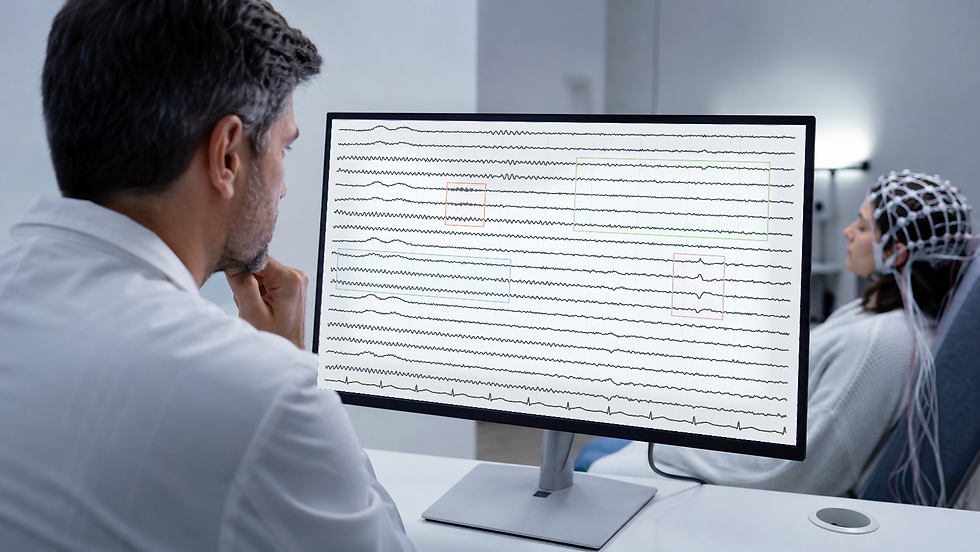
One EEG pattern, spindling excessive beta (SEB), shows particular promise. SEB, first described by Gibbs and Gibbs (1950), manifests as sinusoidal, spindle-like beta activity between 13 and 35 Hz and appears linked to neuroinflammatory states, including traumatic brain injury (TBI) and treatment-refractory psychiatric conditions (Swatzyna et al., 2024). The SEB graphic below is courtesy of Dr. Ronald Swatzyna.

What is the Science?
Neuroinflammation is an immune process centered in the brain and spinal cord, orchestrated primarily by microglia. Graphic © Juan Gaertner/ Shutterstock.com.

These cells account for about 10% of total brain cells and act as first responders when confronted by pathogens, toxins, or trauma (DiSabato, Quan, & Godbout, 2016). While acute activation of microglia helps contain injury, prolonged activation releases cytokines that damage neurons, impair cognition, and may initiate degenerative cascades (Schimmel, Acosta, & Lozano, 2017). This chronic state underlies many clinical symptoms such as fatigue, poor memory, depression, sleep disturbance, and the widely reported “brain fog” observed after COVID-19 infection (University of Birmingham, 2019).
Existing diagnostic tools are limited. Lumbar puncture remains an invasive procedure and yields false negatives in up to 15% of cases (Institute for Quality and Efficiency in Health Care, 2020). MRI may identify demyelinating lesions in multiple sclerosis, but it lacks sensitivity for many inflammatory states (Bechter, 2020). PET can detect microglial activation but requires expensive radioligands (Thelin et al., 2017). Blood biomarkers such as the Banyan Brain Trauma Indicator are validated for TBI but not general neuroinflammation (University of Florida Research, 2023). Brain biopsy, while definitive, is too invasive for widespread use. EEG, however, is non-invasive, repeatable, and provides a direct window into brain physiology, capturing inflammatory effects on neuronal networks in real time (Woodward et al., 2020).
What Did They Study?
The study focused on spindling excessive beta (SEB), an EEG phenotype distinguished from typical irregular beta activity by its sinusoidal, spindled morphology. SEB was first reported by Gibbs, Lorimer, and Gibbs (1950) and later linked to barbiturate toxicity (Gibbs & Gibbs, 1962).
More recent research has implicated SEB in disorders involving sleep regulation and impulse control (Arns, Swatzyna, Gunkelman, & Olbrich, 2015; Krepel, van Dijk, Sack, Swatzyna, & Arns, 2021). In clinical practice, SEB has been observed in patients with toxic exposures, brain infections, chronic pain, psychiatric illness, and especially TBI. The authors hypothesized that SEB reflects underlying neuroinflammation regardless of whether its origin is pharmacologic, infectious, or mechanical.
What Did They Do?
The authors retrospectively analyzed EEGs from 1,233 treatment-refractory psychiatric patients, with additional focus on 79 adults with TBI compared against 75 non-TBI adults. Recordings followed standardized protocols: 19-channel 10–20 electrode placements, linked-ear references, impedance < 5 kΩ, and 20–40 minutes of resting-state acquisition. Experienced analysts removed artifacts, and a board-certified electroencephalographer reviewed all studies, remaining blind to the diagnosis.
Two statistical approaches characterized the SEB distribution:
Auto-Contractive Map (Auto-CM) with Minimum Spanning Tree (MST). Auto-CM is a type of artificial neural network designed not for prediction but for knowledge discovery. It constructs a weighted graph of all variable relationships and reduces it to a semantic connectivity map. This map can then be simplified using MST, which identifies the smallest set of edges needed to connect all nodes, highlighting the most meaningful associations (Buscema & Grossi, 2008; Buscema, Grossi, Snowdon, & Antuono, 2008). Stability was tested through resampling, with topological consistency exceeding 98%.
Binary logistic regression. This model evaluated which electrode sites predicted TBI versus non-TBI group membership. Right-lateralized electrodes (T4, Fp2) were identified as strong risk markers, while midline (Fz) and left temporal (T5) activity acted as protective features. Sensitivity reached 87.3% and specificity 82.7%, yielding an overall classification accuracy of 85.1%.
What Did They Find About Spindling Excessive Beta?
SEB was present in 25.1% of the total cohort. Children and adolescents showed prevalence rates around 20–22%, while adults had a significantly higher rate at 31.5%. Across all groups, females exhibited slightly higher prevalence than males, a pattern consistent with higher rates of anxiety and panic disorders in women (Swatzyna et al., 2024).
The distinction between TBI and non-TBI groups was striking. SEB prevalence reached nearly 78% in adults with TBI compared to 31.5% in adults without TBI. Topographically, non-TBI patients showed SEB concentrated in the frontal and central regions, whereas TBI patients exhibited SEB shifted into temporal regions. These findings align with the known biomechanical vulnerability of the temporal lobes during head trauma (Schimmel et al., 2017).
What is the Impact?
These findings suggest that EEG, and specifically SEB, can serve as a clinically valuable biomarker for neuroinflammation. For psychiatrists and neurologists, this means moving beyond symptom-driven diagnoses and integrating physiological evidence into treatment planning. For instance, selective serotonin reuptake inhibitors (SSRIs), though widely prescribed for anxiety, may increase beta activity and inadvertently exacerbate SEB in vulnerable patients (Schmidt, 2023).
Conversely, treatments that improve perfusion and reduce inflammation—guanfacine with NAC, photobiomodulation, and hyperbaric oxygen therapy—appear to reduce SEB, offering a way to both identify and monitor neuroinflammatory states (Gottfried, Schottlender, & Ashery, 2021).
By incorporating EEG biomarkers into psychiatric and neurorehabilitative care, clinicians could triage patients with persistent “brain fog,” refractory anxiety, or post-TBI symptoms more effectively. This approach may represent a paradigm shift toward precision psychiatry, where interventions are guided not just by symptoms but by underlying neurophysiology.
Five Key Take-Aways
1. Chronic neuroinflammation is underdiagnosed and often mimics psychiatric illness.
2. EEG, particularly SEB, is a promising biomarker for inflammation-related brain dysfunction.
3. SEB prevalence is especially high in adults with TBI and shows temporal lobe involvement.
4. Right frontotemporal SEB markers differentiate TBI from non-TBI cases with high sensitivity and specificity.
5. Interventions that enhance cerebral perfusion and reduce inflammation correlate with SEB reduction, suggesting utility for diagnosis and treatment monitoring.

Glossary
auto-contractive map (Auto-CM): an artificial neural network that generates a weighted map of variable relationships for knowledge discovery, not prediction. Used here to reveal EEG topographies.
binary logistic regression: a statistical method used when the outcome has two categories, such as yes/no or present/absent. It predicts the probability of one outcome by modeling the relationship between predictor variables and the log-odds (natural logarithm of the odds) of the event. The results are expressed as odds ratios, showing how each predictor increases or decreases the likelihood of the outcome.
electroencephalography (EEG): a technique that records scalp electrical activity of the brain, used clinically to evaluate cortical function.
extreme delta brush (EDB): an EEG pattern seen in anti-NMDA receptor encephalitis, combining slow delta activity with superimposed spindled beta.
guanfacine (±N-acetylcysteine): a medication acting on α2A adrenergic receptors; when combined with NAC, it may reduce neuroinflammation and SEB.
hyperbaric oxygen therapy (HBOT): a treatment providing oxygen at higher-than-atmospheric pressure, enhancing tissue oxygenation and reducing inflammation.
microglia: resident immune cells of the CNS that coordinate inflammatory responses; beneficial acutely but damaging when chronically activated.
minimum spanning tree (MST): a graph theory algorithm that identifies the shortest path connecting all nodes in a network, highlighting strongest relationships.
neuroinflammation: immune-mediated inflammation in the brain or spinal cord, protective acutely but harmful chronically.
photobiomodulation (PBM): a therapy using red or near-infrared light to stimulate cellular repair and reduce inflammation.
spindling excessive beta (SEB): a sinusoidal, spindle-like beta EEG pattern associated with medication toxicity, impulse control problems, and neuroinflammatory states.
References
Arns, M., Swatzyna, R. J., Gunkelman, J., & Olbrich, S. (2015). Sleep maintenance, spindling excessive beta and impulse control: An RDoC arousal and regulatory systems approach? Neuropsychiatric Electrophysiology, 1(1), 5. https://doi.org/10.1186/s40810-015-0005-4
Bechter, K. (2020). The challenge of assessing mild neuroinflammation in severe mental disorders. Frontiers in Psychiatry, 11, 773. https://doi.org/10.3389/fpsyt.2020.00773
Buscema, M., & Grossi, E. (2008). The semantic connectivity map: An adapting self-organising knowledge discovery method in databases. International Journal of Data Mining and Bioinformatics, 2(4), 362–404. https://doi.org/10.1504/IJDMB.2008.022161
Buscema, M., Grossi, E., Snowdon, D., & Antuono, P. (2008). Auto-Contractive maps: An artificial adaptive system for data mining. An application to Alzheimer disease. Current Alzheimer Research, 5(5), 481–498. https://doi.org/10.2174/156720508785908948
DiSabato, D. J., Quan, N., & Godbout, J. P. (2016). Neuroinflammation: The devil is in the details. Journal of Neurochemistry, 139(S2), 136–153. https://doi.org/10.1111/jnc.13607](https://doi.org/10.1111/jnc.13607
Gibbs, E. L., Lorimer, F. M., & Gibbs, F. A. (1950). Clinical correlates of exceedingly fast activity in the electroencephalogram. Diseases of the Nervous System, 11(11), 323–326.
Gibbs, E. L., & Gibbs, F. A. (1962). Extreme spindles: Correlation of electroencephalographic sleep pattern with mental retardation. Science, 138(3545), 1106–1107. [https://doi.org/10.1126/science.138.3545.1106](https://doi.org/10.1126/science.138.3545.1106)
Gottfried, I., Schottlender, N., & Ashery, U. (2021). Hyperbaric oxygen treatment—from mechanisms to cognitive improvement. Biomolecules, 11(10), 1520. https://doi.org/10.3390/biom11101520
Institute for Quality and Efficiency in Health Care. (2020). What happens during a lumbar puncture (spinal tap)? InformedHealth.org. https://www.ncbi.nlm.nih.gov/books/NBK367574/
Krepel, N., van Dijk, H., Sack, A. T., Swatzyna, R. J., & Arns, M. (2021). To spindle or not to spindle: A replication study into spindling excessive beta as a transdiagnostic EEG feature associated with impulse control. Biological Psychology, 165, 108181. https://doi.org/10.1016/j.biopsycho.2021.108181
Morrow, L. M., Barr, E. A., Grossi, E., Pillai, V. K., Kight, K. A., Wright, E. B., Turner, R. P., & Swatzyna, R. J. (2025). Identifying Neuroinflammation: The Diagnostic Potential of Spindling Excessive Beta in the EEG. Clinical EEG and Neuroscience, 15500594251376475. Advance online publication. https://doi.org/10.1177/15500594251376475
Rhie, S. J., Jung, E.-Y., & Shim, I. (2020). The role of neuroinflammation on pathogenesis of affective disorders. Journal of Exercise Rehabilitation, 16(1), 2–9. https://doi.org/10.12965/jer.2040016.008
Schimmel, S. J., Acosta, S., & Lozano, D. (2017). Neuroinflammation in traumatic brain injury: A chronic response to an acute injury. Brain Circulation, 3(3), 135–142. https://doi.org/10.4103/bc.bc_18_17
Schmidt, C. (2023). Inflammation and brain health. Harvard Medicine Magazine. https://hms.harvard.edu/magazine/aging/inflammation-brain-health
Swatzyna, R. J., Morrow, L., Collins, D., Barr, E., Roark, A., & Turner, R. (2024). Evidentiary significance of routine EEG in refractory cases: A paradigm shift in psychiatry. Clinical EEG and Neuroscience, 56(5), 446–456. https://doi.org/10.1177/15500594241234567
Thelin, E. P., Tajsic, T., Zeiler, F. A., et al. (2017). Monitoring the neuroinflammatory response following acute brain injury. Frontiers in Neurology, 8, 351. https://doi.org/10.3389/fneur.2017.00351)
University of Birmingham. (2019, November 15). Link between inflammation and mental sluggishness shown in new study. ScienceDaily. (https://www.sciencedaily.com/releases/2019/11/191115190337.htm)
University of Florida Research. (2023). FDA approves Banyan Biomarkers’ traumatic brain injury blood test. https://research.ufl.edu/banyanbiomarkers.html
Woodward, K. E., de Jesus, P., & Esser, M. J. (2020). Neuroinflammation and precision medicine in pediatric neurocritical care: Multi-modal monitoring of immunometabolic dysfunction. International Journal of Molecular Sciences, 21(23), 9155. https://doi.org/10.3390/ijms21239155
About the Author

Fred Shaffer earned his PhD in Psychology from Oklahoma State University. He earned BCIA certifications in Biofeedback and HRV Biofeedback. Fred is an Allen Fellow and Professor of Psychology at Truman State University, where has has taught for 50 years. He is a Biological Psychologist who consults and lectures in heart rate variability biofeedback, Physiological Psychology, and Psychopharmacology. Fred helped to edit Evidence-Based Practice in Biofeedback and Neurofeedback (3rd and 4th eds.) and helped to maintain BCIA's certification programs.
Support Our Friends


Comments