
Meeting Neurofeedback's Next-Generation Ethical Challenges

Updated: Jul 12
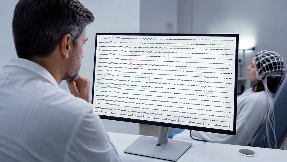
Most neurofeedback providers already bring real care to their work: thoughtful assessment, real-time signal review, artifact control, informed consent, consultation, and outcome monitoring. This article is written to support and celebrate that craft, not to portray the field as careless or uniform. Think of it as a friendly companion for the good practice already underway.

Neurofeedback also carries a distinctive and interesting set of ethical questions, and that is part of what makes it such rewarding work. The path from scalp voltage to a clinical or performance claim passes through electrode placement, reference choice, filtering, artifact handling, feature extraction, normative comparison, thresholding, feedback logic, and human interpretation. Each layer can be beautifully done. Each can also add uncertainty, hide an error, or make a weak inference look more persuasive than it is (Association for Applied Psychophysiology and Biofeedback [AAPB], 2013; Collura et al., 2025; Ros et al., 2020). Noticing those layers is not a criticism of the field; it is simply good stewardship of a powerful tool.
This article focuses on the EEG and the qEEG. It does not claim that every ethical issue discussed here is unique to neurofeedback, nor does it cover every form of neurofeedback or neuromodulation. Where law, regulation, licensure, an employer's policy, or a professional code sets a higher or more specific requirement, that requirement governs.
I am grateful to Donald Moss, Ronald J. Swatzyna, and Frans J. Cronje for conversations that informed my thinking about this rapidly changing area, especially artificial intelligence. I extensively used Dr. Swatzyna and Dr. Barbara Peavey's 2026 Biofeedback Society of Texas ethics presentation. None of my colleagues reviewed or endorsed this article. Any mistakes are exclusively the author's.

Central principle. Keep the client or trainee, the raw physiology, the full context, and accountable human judgment ahead of the tools that summarize them. Everything else in this article is just an elaboration of that one idea.
1. Start by naming the practice context
Neurofeedback is delivered in several materially different contexts, and our ethical language becomes clearer when we simply name the context before we make a claim. Doing so protects both the client and the provider.
Contexts, purposes, and responsibilities
Context or assessment layer | Primary purpose and permissible claims | Core responsibilities |
Clinical assessment or treatment | Address symptoms, impairment, or a diagnosed health condition. Clinical claims stay within the responsible professional's legal scope and competence. | Multimethod assessment, clinical formulation, informed consent, adverse-event monitoring, referral procedures, records, and coordination with other care. |
Performance or educational training | Improve a defined skill or performance outcome in a generally healthy person. Diagnosis or treatment is not implied unless the provider is authorized and the service is actually clinical. | Accurate representation of evidence, safety screening, clear nonclinical boundaries, functional outcome measures, and referral when a health concern emerges. |
Research | Produce generalizable knowledge under an approved protocol. Research participation is not presented as individualized clinical care. | Ethics review where required, protocol fidelity, consent, privacy, adverse-event procedures, appropriate controls, and transparent reporting. CRED-nf is a research design and reporting checklist, not a clinical practice guideline (Ros et al., 2020). |
Use of formal qEEG assessment | Add quantitative summaries or normative comparisons to raw EEG review. | Reviewable raw data, transparent acquisition and artifact procedures, database and method fit, qualified interpretation, and clear limits on diagnostic and treatment-selection claims. |
Some safeguards apply in every context: accurate representation of competence, valid recording, attention to artifact and state, understandable consent, data protection, and a plan for unexpected symptoms. Other duties depend on whether the service is clinical, performance-oriented, or research.
Some neurofeedback approaches do not use formal qEEG, z-scores, or normative databases; the sections on maps, database fit, and quantitative reports apply only when those tools are used, while the raw-signal, artifact, state, consent, supervision, data, and outcome safeguards apply more broadly.
2. Use evidence labels, not a single rhetorical volume
The recommendations below are deliberately labeled. The goal is honesty and humility: an author's preference should never quietly dress itself up as settled consensus. Labeling keeps the conversation open and respectful of the reader's own judgment.
How to read these recommendations
Label | Meaning |
Professional or ethical standard | Explicitly stated in law, regulation, a professional code, or a relevant practice standard. The exact force varies by jurisdiction and profession. |
Evidence-informed risk-management recommendation | Supported by technical, clinical, or ethical evidence, but not necessarily mandated in one universal form. |
Author-proposed conservative clinic policy | A deliberately cautious operational suggestion offered where standards do not prescribe a single method. Clinics may use another model if they can demonstrate equivalent safety, observation, accountability, and legal compliance. |
3. Seven core safeguards
Seven safeguards form the backbone of this framework, and they work best together rather than in isolation. The first is to build raw-signal competence and to know where to find qualified review: anyone who acquires or trains from EEG can learn to verify recording quality, recognize common artifacts and vigilance changes, and sense when a record deserves a more qualified look — a matter of professional guidance joined to evidence-informed risk management.
The second is to let EEG and qEEG inform rather than stand alone whenever a mental or neurodevelopmental question is on the table, since they are wonderful contributors to a picture rather than a solo diagnostic test, a point grounded in professional guidance and biomarker evidence.
The third is to treat candidate biomarkers as good questions rather than verdicts, because a correlation is an invitation to look closer rather than to jump to a diagnosis, cause, prognosis, or automatic training target — an instance of evidence-informed risk management.
The fourth safeguard is to stay curious about competing explanations before interpreting a pattern, because artifact, drowsiness, sleep loss, medication, substances, pain, illness, a past head injury, an environmental exposure, time of day, and recording conditions can all shift the signal; curiosity here is a genuine gift to the client, and it rests on professional guidance and technical evidence.
The fifth is to keep qEEG in a supportive relationship with the raw EEG and the full formulation, so that no map, z-score, classifier, or normative report has to carry decisive weight by itself, again reflecting professional guidance and evidence-informed risk management.
The sixth is to keep accountable clinical authority with qualified people: software may assist within transparent bounds, yet it does not need to silently determine candidacy, diagnosis, treatment target, protocol, or stopping decisions, a stance consistent with broad health-AI guidance.
And the seventh is to design supervision and staffing around continuous observation and prompt help — I recommend that a technician provide neurofeedback training to one client at a time.

4. Raw EEG competence comes first — and it can be a team sport
Competence with the raw EEG is not the same as being a clinical electroencephalographer, and no one needs to carry every level of expertise alone. A fair standard simply distinguishes three levels of responsibility and invites collaboration across them.
Three levels of competence
Level | Minimum competence |
Everyone who acquires or trains from EEG | Verify electrode placement, reference, channel integrity, sampling and filter settings; recognize common eye, muscle, movement, sweat, line, pulse, cable, and electrode artifact; distinguish alert wakefulness from drowsiness well enough to avoid obvious state-trait errors; pause when the signal is invalid or the client is not safely participating. |
Responsible clinician or qualified qEEG reviewer | Judge whether the recording is adequate for the intended use; review in alternate montages and time scales; evaluate normal variants and suspicious focal, generalized, rhythmic, or paroxysmal activity without exceeding the scope of one's health profession; integrate findings with history, symptoms, medication, sleep, and other assessment data; decide when consultation or referral is welcome. |
Medical EEG specialist | Diagnose or formally characterize epileptiform activity, encephalopathy, focal dysfunction, and other medical EEG findings within appropriate credentials and clinical context. |
A provider does not have to perform every level personally. A documented team arrangement with timely access to qualified review addresses the safety need. One caution: a map or automated report is best not relied upon when nobody involved can yet evaluate whether the underlying recording is physiologically plausible.
Current technical guidance for clinical QEEG emphasizes standardized acquisition, visual inspection, artifact management, data selection, and transparent processing (Collura et al., 2025). Routine diagnostic EEG standards offer useful technical context, though they were written for clinical neurophysiology rather than outpatient neurofeedback (Peltola et al., 2023). AAPB and ISNR likewise value competence with the device, physiology, scope, and relevant supervision (AAPB, 2013; International Society for Neuroregulation & Research [ISNR], 2024).
A practical, achievable baseline brings several of these skills together. It means being able to verify the montage, reference, sampling rate, filters, impedances or contact quality, calibration where applicable, and channel integrity, and to inspect unprocessed or minimally processed data and review it in alternate montages when the system permits.
It also means recognizing and, when safe, reproducing common artifacts, distinguishing alert wakefulness from drowsiness and sleep onset, and flagging persistent suspicious activity without assigning a diagnosis outside one's scope.
Just as importantly, it means documenting uncertainty, inviting consultation, and deferring interpretation or training when validity or significance is still an open question.
Practice rule. If the raw record is unavailable, inadequate, or not competently reviewed, the quantitative output is best not given decisive clinical weight. This protects the client and takes pressure off the provider.

5. Knowing when to consult or refer is a strength, not a shortcoming
Referral is never a sign that neurofeedback "failed." It is one of the most ethical things a provider can do, because it protects clients from scope errors and delayed care and often opens the door to the real, treatable driver of their symptoms.
The right action depends on urgency, the provider's own license and competence, and local procedures. A psychiatrist, psychologist, physician, counselor, or other licensed provider may be able to assess and manage a concern directly; another provider may consult or refer.
One of the most helpful mindsets a neurofeedback provider can hold is curiosity about the underlying cause of a presentation. Symptoms that look like anxiety, depression, ADHD, or even psychosis can sometimes arise from substance use, medication effects, a medical condition, a past head injury, or an environmental exposure.
Colleagues such as Swatzyna and Peavey (2026) have made this point vividly for the whole mental-health field: presenting signs and symptoms do not always reveal what is actually happening underneath. For a neurofeedback provider, this means that a thoughtful referral can sometimes unlock relief that no training protocol would have reached on its own.
Immediate emergency or crisis response
Concern | Immediate action | Important limits |
Any current suicidal ideation or recent suicidal behavior | Pause routine neurofeedback and complete or arrange a same-day structured suicide-risk assessment. Active intent, a plan with intent, preparatory behavior, or an inability to maintain safety calls for the clinic's emergency or crisis procedure and the level of care indicated by risk and local law — not an ordinary future referral. In the United States, the 988 Suicide & Crisis Lifeline may be one component of the response, while imminent danger may require emergency services (National Institute of Mental Health [NIMH], n.d.). | A questionnaire score alone does not determine risk. SAFE-T calls for risk and protective factors, suicide inquiry, risk-level determination, intervention, and documentation (Substance Abuse and Mental Health Services Administration [SAMHSA], 2024). |
First or prolonged seizure-like event, sustained altered consciousness, acute focal neurological deficit, or a severe sudden headache with neurological signs | Stop the session and obtain urgent or emergency medical evaluation. | Describe observations rather than diagnosing epilepsy or another disorder unless qualified. Preserve relevant raw data. |
Chest pain, exertional loss of consciousness, severe unexplained shortness of breath, or sustained palpitations with instability | Stop the session and obtain urgent or emergency medical evaluation. | A pulse channel, HRV display, consumer sensor, or EEG artifact is not a diagnostic ECG. |
Prompt consultation or referral
Observation or history | Usual destination | What to document and communicate |
Recurrent unexplained lapses of awareness; new focal neurological symptoms; unexplained cognitive decline or abnormal movements; persistent focal or generalized slowing after artifact, vigilance, medication, and known-history effects have been considered; repeated suspicious sharp, rhythmic, or paroxysmal activity that persists across montages | Neurology or clinical neurophysiology | Objective observations, relevant history, state and medication information, montage and settings, artifact checks, and preserved raw data. Avoid diagnostic labels outside scope. |
Recurrent syncope or presyncope; exertional loss of consciousness; sustained palpitations; chest pain; unexplained dyspnea; family history suggestive of sudden cardiac death; persistent apparent rhythm irregularity after artifact checks; unexpected bradycardia accompanied by symptoms or clinical concern | Primary care, urgent care, or cardiology depending on severity | Syncope evaluation centers on history, examination, and a 12-lead ECG, not qEEG pattern matching (Shen et al., 2017). Asymptomatic sinus bradycardia can be physiologic; context and symptoms matter (Kusumoto et al., 2019). |
Psychosis, mania or hypomania, severe depression, panic, trauma-related symptoms, substance-related instability, or other symptoms that substantially impair safety or function | Assess and manage within the responsible clinician's competence and scope; otherwise consult or refer to an appropriately licensed mental-health professional | Symptoms, onset, duration, impairment, medication and substance factors, risk assessment, and actions taken. qEEG is not the diagnostic basis. |
Loud habitual snoring, witnessed apneas, excessive daytime sleepiness, sleep attacks, marked sleep fragmentation, or persistent insomnia with functional impairment | Primary care or sleep medicine | Sleep history, current sleepiness, medication and substance factors, and whether the recording included drowsiness or sleep. |
Suspected medication interaction, recent medication change with adverse symptoms, intoxication or withdrawal, uncontrolled medical illness, recurrent hypoglycemic symptoms, or another medical concern affecting safe participation | Prescriber, pharmacist, primary care, urgent care, or the relevant specialty | What was taken, dose and timing, recent changes, observed symptoms, vital or device information if valid, and what the provider did. Never direct cessation of prescribed medication outside appropriate authority. |
Nutrition, endocrine, gastrointestinal, chronic pain, or lifestyle concerns without emergency features | Primary care or the relevant licensed professional; an integrative clinician may participate when care is coordinated | Use specific concerns rather than vague labels such as "hormonal imbalance" or "chronic inflammation." Integrative care should complement, not replace or delay, indicated conventional evaluation (National Center for Complementary and Integrative Health [NCCIH], 2021). |
A valuable set of "look a little closer" flags
Several signals suggest that a physiological or environmental contributor may be worth exploring alongside the neurofeedback work. None is alarming on its own; each is simply a prompt to stay curious and, where appropriate, to loop in a medical or specialist colleague (Swatzyna & Peavey, 2026).
An atypical presentation — symptoms that do not fit familiar patterns or carry unusual features — is one such nudge, as is a limited response that shows little movement despite appropriate, well-delivered evidence-based care.
Concurrent physical symptoms, such as unexplained physical complaints or abnormal examination findings alongside the psychological picture, invite the same gentle curiosity, and so does an abrupt onset in which severe symptoms emerge suddenly without a clear psychological precipitant.
Perhaps the most telling prompt of all is an environmental correlation, where symptoms rise and fall with a change of setting or exposure — for example, easing when the person is away from home.
A few findings are best not treated as stand-alone referral triggers
Some observations are simply too nonspecific to send someone along on their own. A supposed "normal-pressure hydrocephalus pattern" inferred from generalized slowing does not qualify, nor does low posterior dominant rhythm amplitude or frequency offered as a test of cerebral perfusion. A PHQ-9 or other questionnaire cutoff is not a referral trigger without clinical assessment and risk inquiry, and neither is a software-generated diagnosis or arrhythmia label drawn from a non-diagnostic signal.
Generalized slowing and posterior rhythm changes are nonspecific.

6. Consider the EEG and qEEG to be contributors, not stand-alone diagnostic tests
A raw EEG can reveal findings that meaningfully change a medical differential, which is genuinely valuable. That is a different thing from diagnosing ADHD, depression, anxiety, PTSD, autism, bipolar disorder, schizophrenia, or a personality disorder.
A qEEG can summarize spectral power, asymmetry, connectivity, phase, source estimates, or database deviations. These metrics are useful for research, measurement, hypothesis generation, and clinical correlation. What they cannot do, on their own, is establish or exclude a mental or neurodevelopmental diagnosis.
The heart of the matter is the gap between group-level association and individual-level decision-making. A feature may differ on average between research samples yet lack the sensitivity, specificity, predictive value, stability, transportability, or incremental validity needed for diagnosis in a particular person. Similar features can appear across diagnoses, in healthy participants, and under fatigue, drowsiness, medication, pain, or artifact (Abi-Dargham et al., 2023; Hammond, 2010).
This is also where the underlying-cause mindset pays off. The same presentation can be shaped by contributors that live outside the EEG entirely — substance use and the medications used to treat it, prescribed psychiatric medications, a mild traumatic brain injury, a finding of a structural deformity of the brain, such as an arachnoid cyst or Chiari malformation, an endocrine or metabolic condition, a nutritional factor, sleep loss, or an environmental exposure (Swatzyna & Peavey, 2026).
Widely used diagnostic frameworks already ask clinicians to consider substance, medical, and environmental factors before assigning a mental-health diagnosis, so a neurofeedback provider who keeps these possibilities in view is simply practicing in good company.
AAPB states that biofeedback is an adjunct and cannot be used alone to make a diagnosis (AAPB, 2013). A responsible diagnostic formulation therefore rests on symptoms, duration, impairment, developmental and medical history, sleep and substance factors, validated measures, differential diagnosis, and the qualified clinician's scope — and would still stand even if the EEG were removed from the chart.
Clinically defensible language
Instead of | Consider |
"The qEEG confirms ADHD." | "This quantitative finding is nonspecific and does not establish or exclude ADHD. It may help guide additional clinical evaluation." |
"This is a depression pattern." | "Similar features have been reported in some research samples, but this individual's finding is not diagnostic and calls for clinical correlation." |
"Your trauma is stored in this brain region." | "This recording cannot localize a psychological experience or determine its cause." |
"The map tells us exactly what to train." | "The map is one helpful data source. A protocol also draws on the raw EEG, the full assessment, the evidence, the person's goals, and a safety plan." |
"The algorithm detected your disorder." | "The software classified a signal feature under a particular model. A qualified professional weighs its validity, limitations, and relevance." |
7. Treat biomarkers as questions, not verdicts
The word biomarker can sound more authoritative than the evidence warrants. A measurable feature can correlate with a disorder yet remain unfit for diagnosis, prognosis, treatment selection, or monitoring — and that is completely okay. It just means the finding is a lead to follow, not a conclusion to announce.
A clinically useful diagnostic biomarker performs at the individual level in the intended population under realistic conditions, with validated thresholds and known error rates. A treatment-selection biomarker indicates that its use improves outcomes compared with not using it.
Psychiatry still lacks robust, broadly validated biomarkers for most individual diagnoses and treatment decisions (Abi-Dargham et al., 2023). This is a shared frontier, not a personal failing.
When a putative EEG biomarker appears, a good next move is to ask a gentle question of it.
Faced with excess theta, one might wonder whether the person is drowsy, sleep-deprived, developmentally younger than the database, taking a sedating medication, or producing eye or movement artifact.
High beta invites a look for facial, temporalis, neck, or scalp EMG, along with a thought about the timing of anxiety, stimulant use, pain, or reference instability. An asymmetry raises its own set of questions: does it replicate, does it persist in alternate montages, and could electrode contact, skull factors, state, or database mismatch explain it?
A connectivity or phase deviation is worth interrogating for stability across clean epochs, preprocessing choices, reference methods, and software implementations. In every one of these cases, no single finding needs to become a protocol target on its own.
A z-score is not the probability of a disease. It is a standardized distance from a reference mean under assumptions about the sample, age range, recording conditions, hardware, preprocessing, artifact handling, and distribution. A value outside a statistical interval can be real yet clinically irrelevant; a value inside the interval can coexist with genuine impairment.
Practice rule. Let a candidate biomarker sharpen the next step — replication, history, testing, consultation, or referral — rather than convert correlation into identity.

8. Interpret the qEEG alongside the raw EEG and the full context
A qEEG is derived from the raw EEG; it is a companion to it, not an independent test. Quantification can compress data, support reproducible measurement, and surface patterns worth reviewing. It can also hide morphology, timing, state transitions, artifacts, and normal variants that decide whether the numbers mean anything. Keeping the two together is where the craft lives.
A defensible and satisfying sequence unfolds in order. It begins by establishing recording quality and verifying settings, then moves to inspecting the raw EEG in appropriate montages and time scales. From there it identifies the person's vigilance and behavioral state, marks, removes, or models artifact using transparent rules, and confirms that enough representative clean data remain. Only then does it make sense to examine quantitative summaries and database comparisons, and finally to integrate the findings with the full clinical or performance formulation.
Reversing this order can quietly invite confirmation bias: a dramatic map appears first, and the reviewer then searches the record for support while overlooking alternatives. Working in order keeps us honest and relaxed.
Keeping the raw data — not only screenshots or a vendor report — helps you and the next reviewer. It lets someone determine which epochs entered the analysis, what artifact decisions were made, what reference and preprocessing were used, which database and software version were applied, and whether the result reproduces after correction.
Current IQCB technical guidance places strong emphasis on these acquisition, inspection, selection, and processing steps (Collura et al., 2025).
Vendor opacity is simply a reason to assign a little less weight to an output. A proprietary system is not automatically invalid, but a report is best not allowed to drive consequential decisions when clinically material details — raw traces, epoch selection, artifact handling, database characteristics, processing parameters, or model limitations — cannot be examined.
CRED-nf supports transparent reporting of signal processing, regulation success, artifact procedures, adverse effects, and clinically meaningful outcomes in neurofeedback experiments (Ros et al., 2020).

9. Expect artifacts, and continuously monitor your client's state
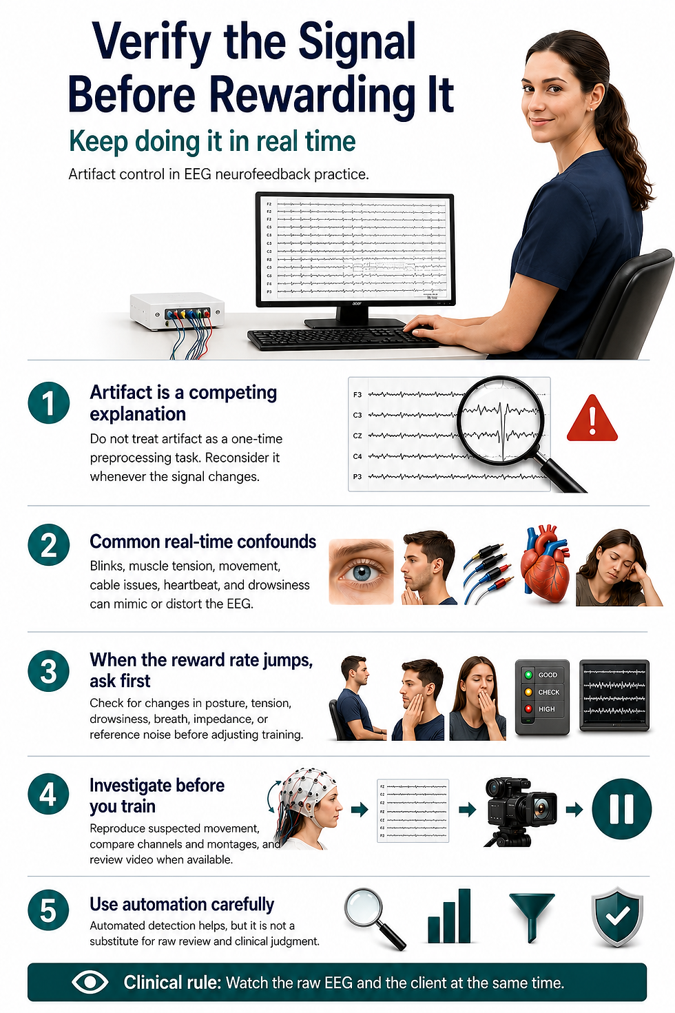
Artifacting is not a one-time housekeeping step finished during preprocessing. It provides a rival explanation, worth reconsidering whenever the signal or reward rate changes. Treating it that way is a mark of expertise, not of doubt.
Blinks and slow eye movements can inflate low frequencies, especially frontally. Jaw, temporalis, neck, and scalp muscles can elevate at higher frequencies. Movement, cable sway, poor contact, bridging, electrode pops, sweat, mains interference, pulse, and reference instability can create spatially convincing noncerebral patterns. Drowsiness can shift alpha and theta in ways that resemble a training effect.
Watch the client or trainee and the raw signal together. When a reward rate jumps, wonder aloud whether the person closed the eyes, tensed the jaw, shifted posture, spoke, swallowed, touched a lead, held the breath, or became drowsy. Check whether contact quality changed, the reference became noisy, or automated correction began removing a different share of the data.
Automated artifact detection is a helpful partner, and it works best when the operator knows what it detects, what it misses, whether it merely withholds feedback or transforms the signal, and how false detections affect learning. Over-aggressive cleaning can remove genuine activity; permissive cleaning can reward artifact. A little familiarity goes a long way.
Pre-recording and pre-session state check
This is also the natural place to stay curious about the wider physiological picture. Many everyday and environmental factors can nudge either the symptoms or the signal, and simply asking about them often reveals a contributor worth addressing or referring (Swatzyna & Peavey, 2026).
A pre-session check
Domain | Ask and document | Typical action |
Sleep and vigilance | Sleep duration and quality, naps, shift work, insomnia, snoring or apnea, current sleepiness | Standardize conditions; pause when drowsy; reschedule or refer when the state is unsafe or unrepresentative. Because sleep and mental health influence each other so strongly, addressing sleep is often a high-value first step. |
Medications and other substances | Prescription and over-the-counter medication, supplements, dose and timing, recent changes, alcohol, cannabis, nicotine, caffeine, and other substances | Consult the prescriber or pharmacist when helpful. Do not independently direct medication cessation. Remember that both substances and the medications used to treat substance use can carry mood and cognitive effects of their own. |
Food, hydration, and nutrition | Time of last meal, a missed usual meal, fluid intake, diabetes, hypoglycemic symptoms, and general dietary patterns | Address immediate safety; document; reschedule or obtain medical care when appropriate. Nutritional factors and blood-sugar swings can influence mood and attention, so a primary-care or nutrition referral is sometimes the kindest next step. |
Illness, pain, and physiology | Fever, infection, migraine, pain flare, endocrine or metabolic symptoms, recent strenuous exercise, and any history of head injury | Defer, modify, or refer according to clinical significance. A history of even mild head injury is worth noting gently, since its effects can emerge over time. |
Environment and setting | Time of day, posture, eyes-open or eyes-closed condition, task, room conditions, and whether symptoms track with a particular living or working environment | Replicate under matched conditions before treating a difference as meaningful. When symptoms clearly ease away from a specific setting, an environmental evaluation may be worth suggesting. |
An EEG samples activity under particular conditions; it is a snapshot, not a fixed portrait of the person. Pharmaco-EEG guidance exists precisely because medications can change vigilance, spectral power, and reactivity (Jobert et al., 2012). Sleep, pain, illness, stress, exercise, caffeine, nicotine, alcohol, cannabis, time of day, hydration, and food intake can also shift state or artifact burden.
A missed meal does not produce one universal EEG pattern. A small 2025 crossover study found no broad behavioral or absolute-power impairment after an 18-hour fast, while reporting a possible frontal-theta change that calls for cautious interpretation (Ávila-Garibay et al., 2025). The practical, non-dramatic point is to document last food intake and symptoms, not to conclude that fasting "caused theta."
10. Keep assessment and planning matched to the context
Clinical treatment
A clinical neurofeedback assessment is richer and more rewarding than qEEG acquisition plus a software report. The responsible clinician gets to know the client's goals, symptoms, functional impairment, developmental and medical history, sleep, medication and substance use, pain, relevant trauma history, current treatment, preferences, and risk factors. Standardized measures fit the actual treatment target.
This is also where a light, curious screen for underlying and contributing causes fits naturally into good care. Without overreaching scope, a clinician can hold open questions about substance use, medication side effects, possible medical or neurological contributors (including past head injuries and structural findings), genetic and metabolic factors, and environmental exposures, and can order or refer for appropriate testing when the picture warrants it.
Frameworks that move from simple observation toward more detailed testing — and that lean on interdisciplinary collaboration — help ensure nothing important is quietly missed (Swatzyna & Peavey, 2026). The aim is not to turn a neurofeedback clinic into a medical workup center; it is to stay alert to the person's whole story and to partner generously with medical and environmental colleagues.
The evaluation is performed or supervised by someone whose license and competence cover the presenting problem. Neurofeedback or qEEG certification does not, by itself, create authority to diagnose or treat outside a professional scope (AAPB, 2013; ISNR, 2024).
A written plan works well when it states the functional target together with the evidence for the proposed approach in this indication and population, and when it names the reasonable alternatives.
It is stronger still when it sets out the expected session range and cost, the outcome measures and a predetermined review point, and the adverse-event monitoring and stop rules, along with the referral and coordination contingencies and — where qEEG was used — how it contributed.
The protocol makes sense from the client's goals, formulation, evidence, and safety profile — even if the qEEG image were removed from the chart.
Performance or educational training
Performance providers do well to define a measurable nonclinical goal, such as sustained attention during a task, recovery after stress, sleep regularity, or a sport-specific outcome. There is no need to convert ordinary variation into a disorder label or to imply that training treats a condition, unless the service is legitimately clinical and within scope.
Safety screening, artifact control, understandable consent, privacy, and referral remain part of the care. When symptoms suggest a medical or diagnosable mental-health condition, the provider warmly explains the limits of the service and encourages consultation with an appropriately licensed professional, as ISNR guidance recognizes (ISNR, 2024).
Research
Research invites a protocol, appropriate ethics review, and consent that distinguish research from care, data governance, adverse-event procedures, and transparent reporting. CRED-nf is especially relevant here because it addresses experimental design, control conditions, regulation success, artifact handling, behavioral outcomes, adverse effects, and reporting (Ros et al., 2020).
A research protocol is best kept from quietly turning into individualized treatment, and clinical services are best not marketed as research to sidestep ordinary professional responsibilities.
When the qEEG and the rest of the assessment disagree
There is no need to give the map automatic priority. A mismatch may reflect artifact, state, database mismatch, unreliable measurement, an incomplete assessment, or a genuine but nonspecific finding. Recheck the raw signal, replicate under matched conditions, broaden the assessment, consult, or gently set the quantitative finding aside. Disagreement is information, not a problem.
11. Let algorithms assist, and keep accountable authority with people
Modern neurofeedback runs on algorithms, and that is a good thing. Filters, transforms, artifact gates, thresholds, reward schedules, adaptive difficulty, source estimates, classifiers, and displays are all computational. The ethical question is not whether an algorithm is present; it is simply whether software might silently control a decision for which a human professional remains accountable.
Where software helps, and where people lead.
Appropriate assistance under human control | Decisions that call for qualified human authority |
Filtering and transforms whose effects are understood | Determining whether a person is an appropriate clinical candidate |
Displaying raw data, trends, and performance | Diagnosing a medical, mental, or neurodevelopmental disorder |
Flagging possible artifact or state changes | Selecting the clinical target and rationale |
Running a clinician-approved contingency | Choosing a new protocol or changing sites or frequency targets for clinical reasons |
Adjusting difficulty or thresholds within explicit, visible, reversible bounds | Deciding that an adverse change is acceptable or that referral is unnecessary |
Logging parameters and changes | Extending treatment or overriding a stop rule without review |
A responsible professional understands the rule, watches its effects, can override it, and documents material changes. Keeping the raw EEG and artifact information visible — rather than watching only the reward display — keeps the human in the loop. Recording software, model, and database versions rounds it out.
Broad WHO and medical-device guidance supports human autonomy, transparency, intended-use clarity, disclosure of limitations, and evaluation of the human-software team (World Health Organization [WHO], 2021; U.S. Food and Drug Administration et al., 2024).
These documents are not neurofeedback practice standards, and FDA device guidance applies only when a product and use fall within the relevant regulatory framework. They are offered here as general design and governance principles, not as direct authority for a particular protocol or staffing model.
Unexpected software behavior is best treated as a quality or safety event: pause, determine what changed, preserve logs, check whether the delivered intervention still matches the approved plan, and report or escalate as appropriate. There is no need to explain an unexplained system change as the client's brain "resisting."

12. Keep technician roles clear, well-supported, and appreciated
AAPB endorses technicians providing clinical biofeedback under the direct supervision of an appropriately trained and licensed clinician. It states that treatment changes are directed by the supervisor and that the supervisor is present at the location where training occurs (AAPB, 2013).
ISNR asks providers to disclose the technician's qualifications, role, and degree of supervision (ISNR, 2024).
A skilled technician contributes enormously — electrode application, recording, coaching, documentation, and implementation of an established plan. To protect both the client and the technician, a few decisions are best kept out of the technician's independent hands: diagnosing or formulating a clinical condition, dismissing suspicious activity as benign, or selecting a clinical target or accepting a software-recommended protocol without review.
In the same spirit, a technician is best not asked to independently change sites, frequency targets, or other clinically material parameters outside delegated bounds, to decide that an adverse change is acceptable, or to decide that referral or escalation is unnecessary.
The supervising professional reviews the assessment and relevant raw EEG, approves the plan and permitted adjustments, defines escalation criteria, remains available as required by the applicable standard and law, and reviews outcomes and adverse events.
A written delegation protocol that names actions permitted independently, actions requiring real-time approval, and actions that are off-limits gives everyone confidence and clarity.
13. Design staffing around observation and response — not a universal slogan
Professional sources support direct supervision, observation, coaching, and accountable changes in treatment. They do not establish a universal one-trainer-to-one-client ratio for every neurofeedback setting, and it is fine to say so plainly.
For clinical neurofeedback, we recommend assigning one technician to one client or to a couple trained together to improve interpersonal synchrony.
A responsible supervisor should be present and accountable as required, clients should understand the staffing model and who is responsible for them, and incidents, near misses, and outcomes are reviewed.
14. Let consent explain uncertainty, supervision, and incidental findings
The step of requesting consent is an opportunity to build trust, not merely a form to complete. Alongside electrode placement and session length, it helps to explain what scalp EEG measures and what qEEG mathematically summarizes, and to be clear that EEG and qEEG are not stand-alone diagnostic tests for mental or neurodevelopmental disorders.
A good consent conversation covers the evidence and uncertainty for the specific indication, protocol, and population; the reasonable alternatives, expected cost and duration, outcome measures, and review points; and the possible adverse experiences the client is warmly welcome to report.
It also specifies who will be present, whether a technician is involved, and who may make protocol changes, describes what adaptive or algorithmic features are active and what their limits are, and explains how incidental or suspicious findings will be reviewed and communicated as well as how data will be stored, shared, processed by vendors, retained, and deleted.
Teach-back can identify misunderstandings: invite the client to explain what the qEEG can and cannot tell them, who makes decisions, what symptoms to report, and when continuation will be revisited.
A neutral incidental-finding statement is often better than reassurance or speculation: "This observation is not diagnostic, but it is worth a look from a professional qualified in medical EEG."
15. Keep an auditable record
A good record aids the next professional caring for your client, helping them to reconstruct what was measured, inferred, delivered, and changed.
At assessment, document the person's state, sleep, medications and other substances, dose timing, food and fluid intake, caffeine and nicotine, time of day, recording conditions, montage, reference, sampling rate, filters, contact quality or impedances, hardware and software versions, artifact procedures, rejected epochs, and the normative database or model used. Preserve the raw EEG and, when applicable, the relationship between raw epochs and quantitative results.
For planning, record the functional target, evidence base, formulation, alternatives considered, reason for the protocol, who approved it, permitted automatic adjustments, stop rules, referral contingencies, and review date.
For each session, record signal quality, observed artifacts, client state, parameters actually delivered, algorithmic changes, coaching, adverse experiences, deviations from plan, and consultation. When a technician conducts the session, identify the technician and supervisor.
"The system adjusted itself" is not quite enough on its own. Version and parameter records make it possible to confirm that a later session still delivered the intervention approved at the outset.

16. Protect neural data and choose vendors thoughtfully
The EEG is not a transcript of thought, but it is sensitive physiological data and deserves respect. Under research conditions, EEG features can even support person identification (Yang et al., 2022). Raw EEG, qEEG features, questionnaires, medication histories, video, and session metadata can together form a detailed longitudinal profile.
It helps to map the actual data flows, minimize collection, separate identifiers where feasible, encrypt storage and transfer, restrict access, set retention and deletion rules, and disclose cloud or vendor processing. The 2026 systematic review of EEG-neurofeedback ethics highlights informed consent, psychological risk, data privacy, vulnerable populations, enhancement, and scientific validity as recurring themes (Ölçüoğlu & Yıldırım, 2026).
Vendor contracts and policies are worth reading for ownership, secondary use, breach response, data location, subcontractors, model training, sale or disclosure, deletion, and export in a usable format.
Obtaining separate authorization for research, teaching, or product-development use — and never making ordinary care contingent on agreeing to unrelated secondary use — keeps trust intact.
Planning for vendor failure or discontinuation ensures clients retain access to their records. A system that blocks access to clinically material raw data, or reserves broad secondary-use rights, is best not allowed to determine care by default.
17. Use stop rules and outcome checkpoints
Setting reassessment and stopping rules before a course begins is a calm, confidence-building habit.
It helps to pause a session whenever the raw signal is invalid, whenever the client becomes drowsy, distressed, medically symptomatic, or unable to participate, or whenever a concerning neurological, cardiac, psychiatric, or physical change appears. The same holds true when software behaves unexpectedly, or when the trainer cannot yet explain what is being reinforced or inhibited.
Stop or substantially revise a course when harms persist, consent is compromised, a needed referral remains incomplete, costs outweigh likely benefit, the protocol has drifted from its rationale, or meaningful progress is absent at the predetermined checkpoint.
Judge outcomes by agreed clinical, functional, educational, or performance measures — not by reward scores or qEEG "normalization" alone. Ask whether attention, sleep, pain, mood, functioning, or performance improved by a meaningful amount, and whether the change can reasonably be linked to training rather than expectancy, concurrent treatment, maturation, practice effects, or regression to the mean.
This is also a natural moment to revisit lifestyle contributors — sleep, nutrition, physical activity, and environment — since gains in those areas can support and sustain the training.
CRED-nf encourages attention to regulation success, behavioral significance, adverse effects, and the relationship between brain regulation and clinical outcomes in research (Ros et al., 2020). Clinics may happily borrow those transparency habits while remembering that CRED-nf is not a practice mandate.
A periodic, practice audit can review raw-signal competence, artifact rates, referrals, protocol changes, staffing and response capacity, adverse events, software versions, consent comprehension, data governance, and outcomes.
EEG neurofeedback often adds modality-specific signal-processing and interpretive layers.
That is reason enough to tailor a few safeguards — without ever claiming that it is inherently more ethical, more dangerous, or more important than any form of EMG, HRV, respiratory, thermal, or other biofeedback.
18. Seven takeaways
Seven ideas capture the spirit of the whole framework. The first is to match claims and duties to the context, remembering that clinical treatment, performance training, research, and formal qEEG assessment each have their own purpose and are not interchangeable.
The second is to build raw-signal competence at the right level, since every operator can grow acquisition, artifact, vigilance, and stop-rule skills while qualified clinical or medical review can be shared across a team.
The third is to let the map inform the work while staying curious about the underlying cause, because EEG and qEEG enrich an evaluation even as a mental or neurodevelopmental diagnosis rests on a full, multimethod picture — one that keeps substance, medical, and environmental contributors in view.
The fourth idea is to treat biomarkers and z-scores as prompts for inquiry, replicating, checking state and artifact, consulting, and correlating before using a finding in a plan.
The fifth is to keep software and quantitative summaries in a supportive relationship with reviewable physiology and accountable people, welcoming automation as an assistant within visible, reversible bounds.
The sixth is to choose supervision and staffing models that can show observation, response, and accountability. A technician should only train one client or couple at a time.
And the seventh is to make the practice auditable, so that clear consent, role boundaries, referral procedures, data governance, stop rules, functional outcomes, and versioned records turn good intentions into observable, sustainable practice.
Glossary
algorithmic clinical authority: the power to make or effectively control a clinical decision. In this framework, accountable authority stays with the responsible qualified person.
artifacts: physiological, environmental, movement-related, or equipment-generated activity that contaminates the EEG or is mistaken for the intended cerebral signal.
automation bias: the tendency to trust a computerized recommendation more than is justified, especially when it looks precise or objective.
bounded assistance: algorithmic or software support that operates within explicit, visible, and reversible limits under human oversight, so that it informs rather than silently controls a clinical decision.
bradycardia: a slow heart rate, generally below 60 beats per minute in adults. It can be a normal finding in physically fit individuals or, when accompanied by symptoms, a sign of a conduction problem
candidate biomarker: a measurable characteristic associated with a biological or clinical process but not yet validated for a specific diagnostic, prognostic, treatment-selection, or monitoring use.
cerebral perfusion: the flow of blood through the brain that delivers oxygen and nutrients to neural tissue. It cannot be validly inferred from the amplitude or frequency of the posterior dominant rhythm.
clinical EEG: EEG obtained and interpreted for medical diagnostic purposes under applicable clinical neurophysiology standards. A neurofeedback recording is not automatically a diagnostic clinical EEG.
CREDnf: the Consensus on the Reporting and Experimental Design of Clinical and Cognitive-Behavioural Neurofeedback studies — a 2020 best-practices checklist that sets standardized guidelines for designing and reporting neurofeedback experiments, covering things like control groups, blinding, pre-registration, and how to document regulation success, in order to improve rigor and reproducibility in the field. focal neurological symptoms: signs or symptoms localized to a specific region of the nervous system, such as one-sided weakness, numbness, visual loss, or speech disturbance, that warrant prompt medical evaluation. hypomania: a distinct period of elevated, expansive, or irritable mood and increased activity that is less severe than mania and does not cause marked functional impairment or psychosis.
incidental finding: an unexpected observation that may have health significance outside the original purpose of the recording and calls for a defined review, communication, and referral process.
intentional dyad: an author-proposed term for two people participating as one defined intervention because their joint interaction is the target. It is not a convenience label for unrelated clients.
montage: the arrangement of EEG channel derivations used to display voltage differences. Different montages can reveal or obscure patterns and artifacts. normal-pressure hydrocephalus pattern: a purported EEG signature of normal-pressure hydrocephalus, typically inferred from generalized slowing. Because generalized slowing is nonspecific, it is not a valid stand-alone basis for diagnosis or referral. palpitations: the subjective awareness of one's own heartbeat, often described as pounding, fluttering, or skipped beats. Sustained palpitations accompanied by instability warrant medical evaluation.
paroxysmal: describing activity or symptoms that begin and end abruptly, such as a sudden burst of EEG activity or an episodic clinical event.
polypharmacy: the concurrent use of multiple medications by one person, which increases the risk of drug interactions and side effects and can alter both the EEG and the clinical presentation.
qEEG: quantitative electroencephalography, the numerical analysis of selected EEG features, sometimes compared with normative data. Its meaning depends on the quality and interpretation of the underlying EEG.
raw EEG: the time-series voltage record before or alongside quantitative summarization.
scope of practice: activities a professional is legally and ethically authorized and competent to perform by virtue of licensure, training, supervision, and jurisdiction. stop rule: a predetermined criterion, set before treatment begins, that specifies when a session or course of training should be paused, revised, or discontinued. syncope: a transient loss of consciousness caused by a temporary reduction in blood flow to the brain, commonly called fainting. Its evaluation centers on history, examination, and a 12-lead ECG rather than qEEG. tachycardia: a fast heart rate, generally above 100 beats per minute in adults, which may be physiologic or a sign of a cardiac or systemic condition.
trait-state distinction: the difference between a relatively enduring characteristic and a temporary condition. A single state-dependent recording is not a stable portrait of a person.
underlying cause: a substance-related, medical, neurological, genetic, or environmental factor that can contribute to — or mimic — a psychiatric presentation, and that may respond to targeted, often interdisciplinary, care.
z-score: a standardized value expressing distance from a reference mean in standard-deviation units. It is not a disease probability or proof that a feature requires treatment.
References
Abi-Dargham, A., Moeller, S. J., Ali, F., DeLorenzo, C., Domschke, K., Horga, G., Jutla, A., Kotov, R., Paulus, M. P., Rubio, J. M., Sanacora, G., Veenstra-VanderWeele, J., & Krystal, J. H. (2023). Candidate biomarkers in psychiatric disorders: State of the field. World Psychiatry, 22(2), 236–262. https://doi.org/10.1002/wps.21078
American Academy of Neurology. (n.d.). Retired guideline notice: Assessment of digital EEG, quantitative EEG, and EEG brain mapping. https://neurologytoday.aan.com/doi/full/10.1212/WNL.49.1.277
Association for Applied Psychophysiology and Biofeedback. (2013). Standards for performing biofeedback. https://aapb.org/Standards_for_Performing_Biofeedback
Ávila-Garibay, A., González-Garrido, A. A., Gómez-Velázquez, F. R., Brofman-
Epelbaum, J. J., Vélez-Pérez, H., Romo-Vázquez, R., & Gallardo-Moreno, G. B. (2025). Fasting and cognitive load-related changes in quantitative EEG measures during an N-back task. International Journal of Psychological Research, 18(1), 59–68. https://doi.org/10.21500/20112084.6690
Collura, T., Cantor, D., Chartier, D., Crago, R., Hartzoge, A., Hurd, M., Kerson, C., Lubar, J., Nash, J., Prichep, L. S., Surmeli, T., Thompson, T., Tracy, M., & Turner, R. (2025). International QEEG Certification Board guideline: Minimum technical requirements for performing clinical quantitative electroencephalography. Clinical EEG and Neuroscience, 56(5), 391–399. https://doi.org/10.1177/15500594241308654
Hammond, D. C. (2010). The need for individualization in neurofeedback: Heterogeneity in QEEG patterns associated with diagnoses and symptoms. Applied Psychophysiology and Biofeedback, 35(1), 31–36. https://doi.org/10.1007/s10484-009-9106-1
Hammond, D. C., Bodenhamer-Davis, G., Gluck, G., Stokes, D. A., Harper, S. H., Trudeau, D. L., MacDonald, M., Lunt, J., & Kirk, L. (2011). Standards of practice for neurofeedback and neurotherapy: A position paper of the International Society for Neurofeedback & Research. Journal of Neurotherapy, 15(1), 54–64. https://doi.org/10.1080/10874208.2010.545760
Hammond, D. C., Walker, J., Hoffman, D., Lubar, J. F., Trudeau, D. L., Gurnee, R., & Horvat, J. (2004). Standards for the use of quantitative electroencephalography (QEEG) in neurofeedback: A position paper of the International Society for Neuronal Regulation. Journal of Neurotherapy, 8(1), 5–27. https://doi.org/10.1300/J184v08n01_02
International Society for Neuroregulation & Research. (2024). Guidelines for practice (ratified July 28, 2024). https://isnr.org/guidelines-for-practice
Jobert, M., Wilson, F. J., Ruigt, G. S. F., Brunovsky, M., Prichep, L. S., Drinkenburg, W. H. I. M., & the IPEG Pharmaco-EEG Guidelines Committee. (2012). Guidelines for the recording and evaluation of pharmaco-EEG data in man: The International Pharmaco-EEG Society (IPEG). Neuropsychobiology, 66(4), 201–220. https://doi.org/10.1159/000343478
Kusumoto, F. M., Schoenfeld, M. H., Barrett, C., Edgerton, J. R., Ellenbogen, K. A., Gold, M. R., Goldschlager, N. F., Hamilton, R. M., Joglar, J. A., Kim, R. J., Lee, R., Marine, J. E., McLeod, C. J., Oken, K. R., Patton, K. K., Pellegrini, C. N., Selzman, K. A., Thompson, A., & Varosy, P. D. (2019). 2018 ACC/AHA/HRS guideline on the evaluation and management of patients with bradycardia and cardiac conduction delay. Circulation, 140(8), e382–e482.
Laborde, S., Mosley, E., & Thayer, J. F. (2017). Heart rate variability and cardiac vagal tone in psychophysiological research: Recommendations for experiment planning, data analysis, and data reporting. Frontiers in Psychology, 8, Article 213. https://doi.org/10.3389/fpsyg.2017.00213
National Center for Complementary and Integrative Health. (2021). Complementary, alternative, or integrative health: What's in a name? https://www.nccih.nih.gov/health/complementary-alternative-or-integrative-health-whats-in-a-name
National Institute of Mental Health. (n.d.). Help for mental illnesses. Retrieved July 12, 2026, from https://www.nimh.nih.gov/health/find-help
Nuwer, M. R. (1997). Assessment of digital EEG, quantitative EEG, and EEG brain mapping: Report of the American Academy of Neurology and the American Clinical Neurophysiology Society. Neurology, 49(1), 277–292. https://doi.org/10.1212/WNL.49.1.277 (Retired guideline; cited for historical context only.)
Ölçüoğlu, R., & Yıldırım, R. V. (2026). Ethical challenges in EEG neurofeedback: A systematic review of gaps, risks, and responsibilities. BMC Medical Ethics, 27, Article 43. https://doi.org/10.1186/s12910-026-01408-4
Peltola, M. E., Leitinger, M., Halford, J. J., Vinayan, K. P., Kobayashi, K., Pressler, R. M., Mindruta, I., Mayor, L. C., Lauronen, L., & Beniczky, S. (2023). Routine and sleep EEG: Minimum recording standards of the International Federation of Clinical Neurophysiology and the International League Against Epilepsy. Epilepsia, 64(3), 602–618. https://doi.org/10.1111/epi.17448
Ros, T., Enriquez-Geppert, S., Zotev, V., Young, K. D., Wood, G., Whitfield-Gabrieli, S., Wan, F., Vuilleumier, P., Vialatte, F., Van De Ville, D., Todder, D., Surmeli, T., Sulzer, J. S., Strehl, U., Sterman, M. B., Steiner, N. J., Sorger, B., Soekadar, S. R., Sitaram, R., … Thibault, R. T. (2020). Consensus on the reporting and experimental design of clinical and cognitive-behavioural neurofeedback studies (CRED-nf checklist). Brain, 143(6), 1674–1685. https://doi.org/10.1093/brain/awaa009
Shen, W.-K., Sheldon, R. S., Benditt, D. G., Cohen, M. I., Forman, D. E., Goldberger, Z. D., Grubb, B. P., Hamdan, M. H., Krahn, A. D., Link, M. S., Olshansky, B., Raj, S. R., Sandhu, R. K., Sorajja, D., Sun, B. C., & Yancy, C. W. (2017). 2017 ACC/AHA/HRS guideline for the evaluation and management of patients with syncope. Circulation, 136(5), e60–e122. https://doi.org/10.1161/CIR.0000000000000499
Substance Abuse and Mental Health Services Administration. (2024). SAFE-T suicide assessment five-step evaluation and triage (Publication No. PEP24-01-036). https://library.samhsa.gov/product/safe-t-suicide-assessment-five-step-evaluation-and-triage/pep24-01-036
Swatzyna, R. J., & Peavey, B. (2026, April 24). Unmasking the underlying cause: An ethical framework for mental health diagnosis [Conference presentation]. Biofeedback Society of Texas.
U.S. Food and Drug Administration, Health Canada, & Medicines and Healthcare products Regulatory Agency. (2024). Transparency for machine learning-enabled medical devices: Guiding principles. https://www.fda.gov/medical-devices/software-medical-device-samd/transparency-machine-learning-enabled-medical-devices-guiding-principles
World Health Organization. (2021). Ethics and governance of artificial intelligence for health: WHO guidance. https://www.who.int/publications/i/item/9789240029200
Yang, Y.-Y., Hwang, A. H.-C., Wu, C.-T., & Huang, T.-R. (2022). Person-identifying brainprints are stably embedded in EEG mindprints. Scientific Reports, 12, Article 17031. https://doi.org/10.1038/s41598-022-21384-0

About the Author
Fred Shaffer earned his PhD in Psychology from Oklahoma State University. He earned BCIA certifications in Biofeedback and HRV Biofeedback. Fred is an Allen Fellow and Professor of Psychology at Truman State University, where he has taught for 50 years. He is a Biological Psychologist who consults and lectures in heart rate variability biofeedback, Physiological Psychology, and Psychopharmacology. Fred helped to edit Evidence-Based Practice in Biofeedback and Neurofeedback (3rd and 4th eds.) and helps to maintain BCIA's certification programs. He is a recipient of AAPB's Distinguished Scientist Award and BFE's Lifetime Impact Award.

Support Our Friends


Comments