
Revolutionary Neuroscience Ideas


The old classroom picture was wonderfully tidy. The brain issued orders, neurons carried them, organs obeyed, and the parasympathetic system cleaned up afterward. That picture helped generations of students, but it mistook a traffic map for the city.

Neuroscience now describes a body built from overlapping networks, local control systems, secretory tissues, glia, immune cells, microbes, and organs that report upstream and sideways (Fornito et al., 2016; Sharkey & Mawe, 2023).
These eight ideas take nothing away from the brain. They simply make the rest of the organism more interesting. The brain remains a powerful coordinator, but it works inside a federation of tissues that sense, predict, communicate, and adapt on several time scales (Benarroch, 1993; Cole et al., 2013; Iadecola, 2017).
One Organ, Several Job Descriptions
A heart looks like a pump because pumping is its most visible job. Its cardiac muscle generates pressure, and pacemaker cells in the sinoatrial node initiate the normal rhythm. Yet the same organ also senses stretch, releases hormones, and contains a local neural network that adjusts its electrical and mechanical behavior (Fedele & Brand, 2020; Ogawa & de Bold, 2014).
The endocrine surprise arrived when atrial tissue produced a rapid natriuretic response in experimental animals. Researchers later identified atrial natriuretic peptide (ANP) and B-type natriuretic peptide (BNP), which help regulate salt, water, vascular tone, and cardiac workload. Clinicians now use circulating natriuretic peptide concentrations to evaluate and monitor heart failure (de Bold et al., 1981; Ogawa & de Bold, 2014).
The neural surprise sits in the intrinsic cardiac nervous system (ICNS). Human hearts contain ganglia and connecting neurons on and within the organ.

These neurons include sensory, local circuit, and efferent elements that integrate signals from the myocardium with sympathetic and parasympathetic input (Armour et al., 1997; Fedele & Brand, 2020).

The ICNS does not replace the sinoatrial node, and it does not turn the heart into a miniature conscious brain. It modulates pacemaking, conduction, contractility, and local reflexes. The wording matters here, because the catchy phrase heart brain can outrun the anatomy (Fedele & Brand, 2020).
The heart is muscle, endocrine tissue, sensory surface, and neural network at the same time. Its identity depends on the question being asked, not on one permanent label (Armour et al., 1997; Ogawa & de Bold, 2014).
Healthcare follows the same logic. A patient with dyspnea may need a pump assessment, a rhythm assessment, a volume assessment, and an autonomic assessment. A single organ can generate several classes of symptoms because several control systems occupy the same tissue (Fedele & Brand, 2020; Ogawa & de Bold, 2014).
The bedside rule is short. Never tell a patient that an organ has only one job. Ask which role is failing, compensating, or being recruited right now (Fedele & Brand, 2020; Ogawa & de Bold, 2014).
Complex Functions Have No Single Address
Older localization diagrams encouraged a real but incomplete intuition: one patch of cortex does one thing. Network science keeps that regional specialization and adds the connections. Functional connectivity describes statistical dependence between regions over time, while structural connectivity describes the axons and pathways that physically link regions (Fornito et al., 2016).

Attention illustrates the change. Voluntary attention recruits dorsal frontoparietal regions, unexpected events recruit ventral attention systems, and the pulvinar helps regulate information flow according to current demands. Sustained attention can be predicted more accurately from whole-brain connectivity than from a single isolated site (Rosenberg et al., 2016; Saalmann et al., 2012).
The salience network, centered on the anterior insula and anterior cingulate cortex, helps decide what deserves processing.

The right fronto-insular cortex also helps switch between the default mode network and the central executive network. This switching function links bodily relevance, attention, and task control (Seeley et al., 2007; Sridharan et al., 2008).

Flexible hubs in the frontoparietal network rapidly change their partners when a new task appears. They distribute instructions and share information with sensory, memory, and motor systems. General intelligence also appears to depend on a distributed network rather than a single anatomical seat (Cole et al., 2013; Dubois et al., 2018).
Autonomic regulation follows the same pattern. The central autonomic network (CAN) links prefrontal, cingulate, insular, amygdalar, hypothalamic, and brainstem regions. It integrates visceral information with goals, emotion, and context before adjusting sympathetic and parasympathetic output (Benarroch, 1993; Thayer & Lane, 2000).

Movement is no longer exempt. The somato-cognitive action network (SCAN) interlaces regions for specific effectors with regions that integrate goals, posture, breathing, arousal, and whole-body action. The discovery helps explain why preparing to speak, reach, or flee changes the heart and breathing before movement begins (Gordon et al., 2023).

Attention, emotional self-regulation, salience, intelligence, and autonomic control do not reside at a single cortical address. They emerge when specialized regions coordinate, compete, and change partners at the right moment (Cole et al., 2013; Seeley et al., 2007).
This changes assessment and treatment. A lesion, electrode, medication, or neurofeedback protocol may alter a network far beyond its immediate target. Network measures can add value, but correlated activity does not prove a direct anatomical connection or a causal pathway (BioSource Faculty, 2025; Fornito et al., 2016).
Every Tissue Has a Voice
The body once looked like a hierarchy of command neurons and obedient tissues. Endocrinology, immunology, and cell biology have replaced that hierarchy with a crowded conversation. Muscle, fat, immune cells, vascular cells, and microbes release signals that change distant organs (Kalluri & LeBleu, 2020; Pedersen & Febbraio, 2012).
Contracting skeletal muscle releases myokines, which are signaling molecules produced by muscle. Interleukin-6 can rise sharply during prolonged exercise, especially when muscle glycogen is low. In that setting, its role differs from the chronic inflammatory signal that clinicians usually associate with elevated interleukin-6 (Pedersen & Febbraio, 2012; Steensberg et al., 2001).

Adipose tissue is also an endocrine organ. Adipocytes release adipokines such as leptin and adiponectin, which influence appetite, insulin sensitivity, inflammation, and energy use. The discovery of leptin helped end the fiction that fat tissue merely stores excess calories (Scherer et al., 1995; Zhang et al., 1994).

Fat communicates with bone through the hormone leptin. In 2002, researchers discovered that fat tissue, via leptin signaling, functions as a major regulator of bone mass.
This fat-to-bone signaling pathway helps explain why changes in body composition, such as significant weight loss or gain, can have profound effects on skeletal health.

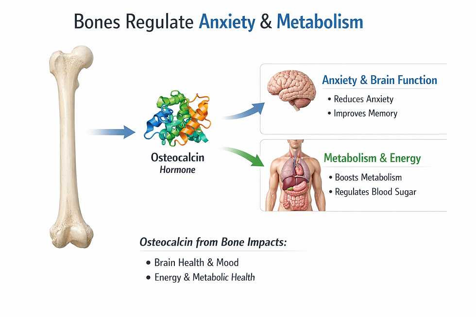
Bone produces the hormone osteocalcin, which has been found to exert effects on multiple organ systems. Osteocalcin influences metabolism and fertility in ways that were entirely unsuspected until relatively recently. Perhaps most remarkably, osteocalcin signals even reach the brain, where research has shown that it reduces anxiety.
This means that the skeleton, long seen as structurally important but biochemically passive, is in fact participating in the regulation of mood and cognitive function through chemical messengers it actively secretes into the bloodstream.
Cells also package proteins, lipids, and nucleic acids inside extracellular vesicles. These membrane-bound parcels can travel through local fluid or circulation and alter recipient cells. Exosomes, one class of extracellular vesicle, can transfer messenger RNA and microRNA between cells (Kalluri & LeBleu, 2020; Valadi et al., 2007).

The microbiome adds another chemical layer. Gut microbes transform food and host compounds into metabolites, including short-chain fatty acids (SCFAs), that influence barrier function and immune regulation. Neural, hormonal, immune, and metabolic routes connect this ecology with the brain and other organs (Cryan et al., 2019; Wastyk et al., 2021).

Human studies link altered gut communities with serious mental illnesses, but the direction of association is unclear. Diet, medication, smoking, sleep, illness severity, and laboratory methods can all reshape the observed community. No stool profile currently diagnoses schizophrenia, bipolar disorder, or major depression (Meehan, 2025; Nguyen et al., 2021; Shaffer, 2026).
Tissues are not silent organs waiting for neural orders. They broadcast hormones, immune messengers, metabolites, and vesicles, then change their behavior when other tissues answer (Kalluri & LeBleu, 2020; Pedersen & Febbraio, 2012).
The lesson for the clinic goes beyond ordering more tests. Take a medication history, diet history, sleep history, infection history, and stress history before interpreting a biomarker or microbiome report. Context often explains the biology better than a single snapshot (Nguyen et al., 2021; Wastyk et al., 2021).
Computation Continues Beyond the Skull
The word computation can mislead if it makes biology sound digital. Here it means transforming inputs into adaptive outputs. By that definition, the nervous system uses distributed control, with several local networks solving immediate problems while exchanging information with the brain and spinal cord (Fornito et al., 2016; Sharkey & Mawe, 2023).
The enteric nervous system (ENS) contains extensive networks of neurons and glia within the gut wall. It coordinates motility, secretion, blood flow, barrier function, and local defense through circuits that can operate without moment-to-moment cortical instructions. Central autonomic pathways still modulate those circuits, so independence and isolation are not the same thing (Furness et al., 2014; Sharkey & Mawe, 2023).

Enteroendocrine cells provide rapid sensory access to this system. They detect nutrients and release hormones, and some form fast synaptic-like connections with vagal sensory fibers in animal models. This pathway gives the brain quick information about the intestinal environment without sending microbes or gut serotonin into the brain (Kaelberer et al., 2018; Sharkey & Mawe, 2023).
The heart uses a similar arrangement. Intracardiac ganglia integrate local mechanical and chemical information with signals arriving from outside the organ. Local reflexes can adjust regional cardiac behavior before a conscious sensation ever appears (Armour et al., 1997; Fedele & Brand, 2020).
Distributed control does not mean that the gut or heart thinks like a person. It means that evolution placed useful decision rules close to the variables they regulate. A local controller can act quickly, reduce transmission demands, and continue operating when higher centers are distracted or impaired (Furness et al., 2014; Sharkey & Mawe, 2023).
The organism works less like a chief executive issuing commands and more like a federation. Local systems handle local problems, while the brain coordinates conflicts, priorities, memory, and long-range prediction (Benarroch, 1993; Sharkey & Mawe, 2023).
This picture rewards clinicians who think across systems. Syncope, functional gastrointestinal symptoms, panic, arrhythmia, and dysautonomia can cross traditional specialty borders. The first task is not to decide which organ is guilty. It is to map the loops that may be amplifying one another (Benarroch, 1993; Sharkey & Mawe, 2023).
Evolution Rewires, It Does Not Stack Floors
The triune brain model pictured a reptilian core, a mammalian emotional layer, and a rational human cortex added in sequence. It caught on because it was easy to remember. Comparative neuroscience shows that it is not an accurate account of vertebrate brain evolution (Cesario et al., 2020).

Vertebrates share ancient forebrain divisions and related cell types. Evolution changes proportions, connectivity, developmental programs, and cellular specializations. It does not preserve three separately operating brains inside one skull (Cesario et al., 2020; Tosches et al., 2018).
The pallium is the vertebrate forebrain territory that gives rise to the cerebral cortex in mammals and related structures in other vertebrates. Single-cell transcriptomics in reptiles reveals both conserved cell classes and lineage-specific innovations. Those findings support branching modification, not a staircase of newer layers sitting on older minds (Tosches et al., 2018).
The cortex is not a detached late arrival that simply overrules primitive centers. Cortical and subcortical regions form recurrent loops, and both participate in perception, learning, action, emotion, and homeostasis. A threat response is not a reptile seizing control from a human executive (Cesario et al., 2020; Fornito et al., 2016).
Evolution did not place a rational mammalian brain atop an emotional reptilian brain. It repeatedly remodeled shared circuits, changed their connections, and built new capacities from old developmental materials (Cesario et al., 2020; Tosches et al., 2018).
Patient education improves when we retire the moral drama. Saying that a primitive brain hijacked a rational brain can make fear or anger sound like personal failure. Describing a threat network that learned too well is more accurate and often more compassionate (Cesario et al., 2020).
The better move is plain, specific language. Name the amygdala, periaqueductal gray, prefrontal cortex, autonomic output, memory, and context when those mechanisms matter. Leave the imaginary reptile out of the consultation (Cesario et al., 2020).
Parasympathetic Physiology Has More Than One Gear
Rest-and-digest is a useful doorway, not a complete map. Parasympathetic pathways support digestion, secretion, cardiac slowing, and recovery. They also participate in rapid shifts that prepare attention, movement, social behavior, and defensive immobility (Roelofs, 2017; Thayer & Lane, 2000).
At the onset of exercise or urgent action, heart rate often rises first through vagal withdrawal, which removes part of the ongoing parasympathetic restraint on the sinoatrial node. Sympathetic activation becomes more influential as intensity rises. The parasympathetic system therefore helps create acceleration by releasing a brake, not only by producing calm (White & Raven, 2014).
Heart rate variability (HRV) captures beat-to-beat variation in cardiac timing. At rest, several HRV measures reflect respiratory and vagal influences, and higher flexible variability often tracks stronger prefrontal-amygdala regulation. HRV is not a direct measure of character, safety, or overall vagal health because posture, breathing, age, medication, rhythm, and measurement choices matter (Sakaki et al., 2016; Thayer et al., 2009).

Freezing adds another twist. Defensive freezing can combine motor inhibition and heart-rate deceleration with increased sensory sampling and readiness for action. It is an active defensive mode, not simple relaxation (Roelofs, 2017).
Under severe or inescapable threat, some people report tonic immobility, an involuntary state of profound movement inhibition. Laboratory work with trauma-exposed participants has linked these reports to restricted body sway and altered cardiac regulation. In human clinical writing, tonic immobility is usually more precise than death-feigning, a term drawn mainly from animal behavior (Volchan et al., 2011).
The polyvagal theory has given clinicians an influential vocabulary for safety, social engagement, mobilization, and shutdown. Its broad emphasis on bodily state and relationship has clinical appeal. Reviewers have challenged several of its anatomical and evolutionary premises, so the model should not be presented as settled neuroanatomy (Grossman, 2023; Neuhuber & Berthoud, 2022; Porges, 2022).

Parasympathetic activity is not a biological parking brake. It can restrain the heart, release that restraint for action, shape attention, and participate in defensive stillness, depending on the circuit and the moment (Roelofs, 2017; White & Raven, 2014).
Trauma-informed care follows from this. Immobility does not prove calm, agreement, or lack of effort. A quiet body can contain intense threat physiology, and the safest intervention may begin with orientation, choice, and paced reconnection rather than an order to relax (Roelofs, 2017; Volchan et al., 2011).
The Synapse Is a Doorway, Not the Whole Street
The classical chemical synapse remains indispensable. An action potential reaches an axon terminal, calcium enters, vesicles release a transmitter, and receptors change the target cell. Ions carry the electrical events, while neurotransmitters and neuromodulators shape changes that can last from milliseconds to much longer periods (Bear et al., 2020; Breedlove & Watson, 2023).

Dale's old principle was often taught as one neuron, one transmitter. Modern work shows co-transmission, in which one neuron releases more than one signaling substance. The mix can vary by terminal, vesicle, firing pattern, and target (Svensson et al., 2019).

Many signals also escape the narrow synaptic cleft. Volume transmission occurs when a transmitter diffuses through extracellular fluid and reaches receptors at some distance. Axonal varicosities can release chemicals along an axon like a drip line rather than only at a terminal button (Coggan et al., 2005; Breedlove & Watson, 2023).

Communication can run backward. In retrograde signaling, a postsynaptic cell releases a messenger that changes transmitter release from the presynaptic cell. Endocannabinoids provide a well-established example in the hippocampus (Wilson & Nicoll, 2001).

Neurons can also communicate through electrical synapses. Gap junction channels allow ions and small molecules to pass directly between cells, often in both directions. Their speed helps groups of neurons synchronize activity (Connors & Long, 2004).

Some neurons act as endocrine cells. A neurohormone is released by a neuron into the circulation or portal blood to act at distant targets. Hypothalamic neurons that release oxytocin, vasopressin, or releasing hormones blur the textbook boundary between nervous and endocrine communication (Breedlove & Watson, 2023).
Neural communication is not a row of private wires. It includes synapses, diffuse chemical fields, backward messages, peptide signals, hormones, and direct ionic bridges (Bear et al., 2020; Connors & Long, 2004; Svensson et al., 2019).
This complexity should temper simple chemical-imbalance stories. A drug can change receptors, transporters, release probability, network rhythms, and feedback loops across several tissues. One molecule rarely maps neatly onto one mood, thought, or diagnosis (Bear et al., 2020; Svensson et al., 2019).
Neurons Share the Work
Neurons generate spikes and long-range signals, but they do not process information alone. Astrocytes regulate extracellular ions, remove neurotransmitters, supply metabolic support, shape synapse formation, and help match local blood flow to neural activity. Their processes surround many synapses and contribute to what researchers call the tripartite synapse (Allen & Lyons, 2018; Khakh & Sofroniew, 2015).

Microglia are the resident immune cells of the central nervous system. During development, they help remove selected synapses, and their signaling continues to influence repair, inflammation, and circuit function across life. Excessive or mistimed microglial activity can alter a network without killing its neurons outright (Allen & Lyons, 2018; Paolicelli et al., 2011).

Oligodendrocytes make central myelin, which changes conduction speed and the timing of network communication. In mice, blocking new oligodendrocyte formation disrupts the acquisition of a new motor skill. Learning can therefore require changes in the insulation and timing of axons, not only changes at synapses (McKenzie et al., 2014).

Blood vessels also join the calculation. The neurovascular unit includes neurons, glia, endothelial cells, pericytes, and vascular smooth muscle. These elements coordinate blood flow, barrier function, and metabolic delivery, which is why a functional MRI signal reflects vascular responses coupled to neural activity rather than direct recordings of thought (Iadecola, 2017).

The boldest claims here deserve restraint. Astrocytes clearly sense neural activity and regulate the synaptic environment. Whether they routinely release transmitters as a physiological signaling system, called gliotransmission, remains contested because different methods and preparations produce different answers (Fiacco & McCarthy, 2018; Savtchouk & Volterra, 2018).
Neurons remain essential, but they are not sole proprietors of information processing. Glia and vascular cells control the chemistry, energy, timing, pruning, insulation, and blood supply that make neural computation possible (Allen & Lyons, 2018; Iadecola, 2017).
The clinical payoff here is real. Neurodegeneration, epilepsy, psychiatric illness, trauma, and vascular disease can involve failures in neuron-glia-vascular cooperation. A normal-looking neuron does not guarantee a healthy circuit if its support system is inflamed, poorly myelinated, metabolically strained, or badly perfused (Allen & Lyons, 2018; Iadecola, 2017).
The more accurate sentence is not neurons are irrelevant. It is neurons are not in sole charge. That small edit changes the research question, the treatment target, and the story we tell patients (Allen & Lyons, 2018).
From Command Center to Conversation
The revolutionary change is not one new structure or molecule. It is a change in explanatory style. Neuroscience is moving from isolated parts and one-way commands toward nested networks that exchange information across cells, organs, and time scales (Fornito et al., 2016; Sharkey & Mawe, 2023).
That model leaves room for specialization. The sinoatrial node still paces the heart, neurons still fire action potentials, and cortical regions still make distinctive contributions. The difference is that no component acts meaningfully outside the system that supports, constrains, and interprets it (Allen & Lyons, 2018; Fedele & Brand, 2020).
The nervous system is less a throne than a conversation. Health depends on whether its participants can exchange signals, shift roles, recover flexibility, and coordinate action without one loop drowning out the rest (Benarroch, 1993; Thayer et al., 2009).
For healthcare professionals, this is not decorative theory. It changes history taking, patient education, interpretation of biomarkers, and the design of interventions. Treat the loop in front of you, but keep asking which other loops are feeding it (Benarroch, 1993; Sharkey & Mawe, 2023).
Five Key Neuroscience Takeaways
1. A biological structure can hold several identities at once. The heart is muscle, endocrine tissue, sensory tissue, and a locally innervated control system, so assessment should match the role under strain.
2. Integrative functions belong to networks. Attention, salience, autonomic control, self-regulation, and intelligence depend on dynamic cooperation among specialized regions, not on a single permanent center.
3. The body communicates in every direction. Tissues release hormones, cytokines, metabolites, and vesicles, while local nervous systems in the gut and heart transform information before the brain receives it.
4. Familiar teaching metaphors need guardrails. Rest-and-digest, the triune brain, the chemical synapse, and the neuron-as-boss can introduce a topic, but they become misleading when presented as complete physiology.
5. Clinical care improves when it targets coordination. Ask how neural, endocrine, immune, microbial, glial, vascular, and behavioral loops interact, then choose interventions that restore flexible regulation rather than chasing a single master switch.
Glossary
adipokine: a signaling molecule released by adipose tissue that can influence metabolism, appetite, vascular function, and inflammation.
astrocyte: a star-shaped glial cell that regulates ions, neurotransmitter clearance, metabolism, synapse development, and local blood flow.
central autonomic network (CAN): interconnected forebrain, limbic, hypothalamic, and brainstem regions that integrate bodily information and coordinate autonomic output.
central executive network: a large-scale network centered on lateral prefrontal and parietal regions that supports working memory, attention, and goal-directed control.
co-transmission: the release of more than one signaling substance by a neuron, with release depending on its terminals and activity pattern.
default mode network: a large-scale network active during internally directed cognition, autobiographical reflection, and spontaneous thought.
distributed control: a control architecture in which local systems transform information and regulate local variables while coordinating with higher and neighboring systems.
electrical synapse: a gap-junction connection that permits rapid passage of ions and small molecules between cells, often in both directions.
enteric nervous system (ENS): the networks of neurons and glia in the gut wall that control digestion, secretion, blood flow, barrier function, and local defense.
extracellular vesicle: a membrane-bound particle released by a cell that carries proteins, lipids, or nucleic acids to other cells.
frontoparietal network: a flexible control network linking lateral prefrontal and parietal regions that changes its connections according to task demands.
functional connectivity: statistical dependence or correlated activity between regions over time, which does not by itself prove a direct anatomical connection.
gliotransmission: the proposed regulated release of signaling molecules from glia to influence neurons or other glia, a physiological role that remains debated.
heart rate variability (HRV): beat-to-beat variation in cardiac timing that reflects several interacting influences, including respiration and cardiac vagal modulation.
intrinsic cardiac nervous system (ICNS): ganglia and connecting neurons within the heart that integrate local sensory information with extrinsic autonomic signals.
microglia: resident immune cells of the central nervous system that survey tissue, shape synapses, and participate in inflammation and repair.
myokine: a signaling molecule released by skeletal muscle, especially during contraction, that can act locally or on distant tissues.
natriuretic peptide: a cardiac hormone, including ANP and BNP, that helps regulate fluid volume, vascular tone, and cardiac workload.
neurohormone: a chemical messenger released by a neuron into blood or portal circulation to act on distant targets.
neurovascular unit: the interacting neurons, glia, endothelial cells, pericytes, and vascular muscle that regulate cerebral blood flow and barrier function.
oligodendrocyte: a central nervous system glial cell that forms myelin around axons and helps regulate conduction timing.
pallium: the dorsal forebrain territory that produces the mammalian cerebral cortex and related structures in other vertebrates.
polyvagal theory: a clinical and theoretical model linking vagal pathways with safety, social engagement, mobilization, and shutdown; several anatomical and evolutionary claims remain disputed.
retrograde signaling: communication in which a postsynaptic cell sends a messenger backward to alter presynaptic release.
salience network: a network centered on the anterior insula and anterior cingulate cortex that identifies behaviorally relevant internal and external events and helps allocate processing resources.
short-chain fatty acid (SCFA): a microbial fermentation product, such as acetate, propionate, or butyrate, that acts as a fuel and signaling molecule.
somato-cognitive action network (SCAN): interconnected motor-region nodes that integrate goals, posture, breathing, cardiovascular state, and whole-body action.
structural connectivity: the physical axonal projections and pathways that link nervous system regions.
tonic immobility: an involuntary defensive state marked by profound movement inhibition under severe or inescapable threat.
tripartite synapse: a synaptic unit that includes presynaptic and postsynaptic neuronal elements plus surrounding astrocytic processes.
triune brain model: the inaccurate idea that human brains contain separately operating reptilian, emotional mammalian, and rational cortical layers added in sequence.
vagal withdrawal: a reduction in ongoing cardiac parasympathetic restraint that allows heart rate to rise rapidly during action or exercise.
volume transmission: extrasynaptic signaling in which a chemical messenger diffuses through extracellular fluid to reach receptors beyond a single synaptic cleft.
References
Allen, N. J., & Lyons, D. A. (2018). Glia as architects of central nervous system formation and function. Science, 362(6411), 181-185. https://doi.org/10.1126/science.aat0473
Armour, J. A., Murphy, D. A., Yuan, B. X., Macdonald, S., & Hopkins, D. A. (1997). Gross and microscopic anatomy of the human intrinsic cardiac nervous system. The Anatomical Record, 247(2), 289-298. https://doi.org/10.1002/(SICI)1097-0185(199702)247:2<289::AID-AR15>3.0.CO;2-L
Bear, M. F., Connors, B. W., & Paradiso, M. A. (2020). Neuroscience: Exploring the brain (4th enhanced ed.). Jones & Bartlett Learning.
Benarroch, E. E. (1993). The central autonomic network: Functional organization, dysfunction, and perspective. Mayo Clinic Proceedings, 68(10), 988-1001. https://doi.org/10.1016/S0025-6196(12)62272-1
BioSource Faculty. (2024a, August 7). Neuroscience breakthroughs since graduate school - Part 4: Neurons. BioSource Software.
BioSource Faculty. (2024b, November 7). Neuroscience breakthroughs since graduate school - Part 6: Stress. BioSource Software.
BioSource Faculty. (2025, April 29). A review of the networks trained by neurofeedback. BioSource Software.
Breedlove, S. M., & Watson, N. V. (2023). Behavioral neuroscience (10th ed.). Oxford University Press.
Cesario, J., Johnson, D. J., & Eisthen, H. L. (2020). Your brain is not an onion with a tiny reptile inside. Current Directions in Psychological Science, 29(3), 255-260. https://doi.org/10.1177/0963721420917687
Coggan, J. S., Bartol, T. M., Esquenazi, E., Stiles, J. R., Lamont, S., Martone, M. E., Berg, D. K., Ellisman, M. H., & Sejnowski, T. J. (2005). Evidence for ectopic neurotransmission at a neuronal synapse. Science, 309(5733), 446-451. https://doi.org/10.1126/science.1108239
Cole, M. W., Reynolds, J. R., Power, J. D., Repovs, G., Anticevic, A., & Braver, T. S. (2013). Multi-task connectivity reveals flexible hubs for adaptive task control. Nature Neuroscience, 16(9), 1348-1355. https://doi.org/10.1038/nn.3470
Connors, B. W., & Long, M. A. (2004). Electrical synapses in the mammalian brain. Annual Review of Neuroscience, 27, 393-418. https://doi.org/10.1146/annurev.neuro.26.041002.131128
Cryan, J. F., O'Riordan, K. J., Cowan, C. S. M., Sandhu, K. V., Bastiaanssen, T. F. S., Boehme, M., Codagnone, M. G., Cussotto, S., Fulling, C., Golubeva, A. V., Guzzetta, K. E., Jaggar, M., Long-Smith, C. M., Lyte, J. M., Martin, J. A., Molinero-Perez, A., Moloney, G., Morelli, E., Morillas, E., ... Dinan, T. G. (2019). The microbiota-gut-brain axis. Physiological Reviews, 99(4), 1877-2013. https://doi.org/10.1152/physrev.00018.2018
de Bold, A. J., Borenstein, H. B., Veress, A. T., & Sonnenberg, H. (1981). A rapid and potent natriuretic response to intravenous injection of atrial myocardial extract in rats. Life Sciences, 28(1), 89-94. https://doi.org/10.1016/0024-3205(81)90370-2
Dubois, J., Galdi, P., Paul, L. K., & Adolphs, R. (2018). A distributed brain network predicts general intelligence from resting-state human neuroimaging data. Philosophical Transactions of the Royal Society B: Biological Sciences, 373(1756), 20170284. https://doi.org/10.1098/rstb.2017.0284
Fedele, L., & Brand, T. (2020). The intrinsic cardiac nervous system and its role in cardiac pacemaking and conduction. Journal of Cardiovascular Development and Disease, 7(4), 54. https://doi.org/10.3390/jcdd7040054
Fiacco, T. A., & McCarthy, K. D. (2018). Multiple lines of evidence indicate that gliotransmission does not occur under physiological conditions. The Journal of Neuroscience, 38(1), 3-13. https://doi.org/10.1523/JNEUROSCI.0016-17.2017
Fornito, A., Zalesky, A., & Bullmore, E. T. (2016). Fundamentals of brain network analysis. Academic Press.
Furness, J. B., Callaghan, B. P., Rivera, L. R., & Cho, H. J. (2014). The enteric nervous system and gastrointestinal innervation: Integrated local and central control. Advances in Experimental Medicine and Biology, 817, 39-71. https://doi.org/10.1007/978-1-4939-0897-4_3
Gordon, E. M., Chauvin, R. J., Van, A. N., Rajesh, A., Nielsen, A., Newbold, D. J., Lynch, C. J., Seider, N. A., Krimmel, S. R., Scheidter, K. M., Monk, J., Miller, R. L., Metoki, A., Montez, D. F., Zheng, A., Elbau, I., Madison, T., Nishino, T., Myers, M. J., ... Dosenbach, N. U. F. (2023). A somato-cognitive action network alternates with effector regions in motor cortex. Nature, 617(7960), 351-359. https://doi.org/10.1038/s41586-023-05964-2
Grossman, P. (2023). Fundamental challenges and likely refutations of the five basic premises of the polyvagal theory. Biological Psychology, 180, 108589. https://doi.org/10.1016/j.biopsycho.2023.108589
Iadecola, C. (2017). The neurovascular unit coming of age: A journey through neurovascular coupling in health and disease. Neuron, 96(1), 17-42. https://doi.org/10.1016/j.neuron.2017.07.030
Kaelberer, M. M., Buchanan, K. L., Klein, M. E., Barth, B. B., Montoya, M. M., Shen, X., & Bohórquez, D. V. (2018). A gut-brain neural circuit for nutrient sensory transduction. Science, 361(6408), eaat5236. https://doi.org/10.1126/science.aat5236
Kalluri, R., & LeBleu, V. S. (2020). The biology, function, and biomedical applications of exosomes. Science, 367(6478), eaau6977. https://doi.org/10.1126/science.aau6977
Khakh, B. S., & Sofroniew, M. V. (2015). Diversity of astrocyte functions and phenotypes in neural circuits. Nature Neuroscience, 18(7), 942-952. https://doi.org/10.1038/nn.4043
McKenzie, I. A., Ohayon, D., Li, H., de Faria, J. P., Emery, B., Tohyama, K., & Richardson, W. D. (2014). Motor skill learning requires active central myelination. Science, 346(6207), 318-322. https://doi.org/10.1126/science.1254960
Meehan, Z. (2025, January 23). 5-Min Science: The gut microbiome in serious mental illnesses. BioSource Software.
Neuhuber, W. L., & Berthoud, H. R. (2022). Functional anatomy of the vagus system: How does the polyvagal theory comply? Biological Psychology, 174, 108425. https://doi.org/10.1016/j.biopsycho.2022.108425
Nguyen, T. T., Hathaway, H., Kosciolek, T., Knight, R., & Jeste, D. V. (2021). Gut microbiome in serious mental illnesses: A systematic review and critical evaluation. Schizophrenia Research, 234, 24-40. https://doi.org/10.1016/j.schres.2019.08.026
Ogawa, T., & de Bold, A. J. (2014). The heart as an endocrine organ. Endocrine Connections, 3(2), R31-R44. https://doi.org/10.1530/EC-14-0012
Paolicelli, R. C., Bolasco, G., Pagani, F., Maggi, L., Scianni, M., Panzanelli, P., Giustetto, M., Ferreira, T. A., Guiducci, E., Dumas, L., Ragozzino, D., & Gross, C. T. (2011). Synaptic pruning by microglia is necessary for normal brain development. Science, 333(6048), 1456-1458. https://doi.org/10.1126/science.1202529
Pedersen, B. K., & Febbraio, M. A. (2012). Muscles, exercise and obesity: Skeletal muscle as a secretory organ. Nature Reviews Endocrinology, 8(8), 457-465. https://doi.org/10.1038/nrendo.2012.49
Porges, S. W. (2022). Polyvagal theory: A science of safety. Frontiers in Integrative Neuroscience, 16, 871227. https://doi.org/10.3389/fnint.2022.871227
Roelofs, K. (2017). Freeze for action: Neurobiological mechanisms in animal and human freezing. Philosophical Transactions of the Royal Society B: Biological Sciences, 372(1718), 20160206. https://doi.org/10.1098/rstb.2016.0206
Rosenberg, M. D., Finn, E. S., Scheinost, D., Papademetris, X., Shen, X., Constable, R. T., & Chun, M. M. (2016). A neuromarker of sustained attention from whole-brain functional connectivity. Nature Neuroscience, 19(1), 165-171. https://doi.org/10.1038/nn.4179
Saalmann, Y. B., Pinsk, M. A., Wang, L., Li, X., & Kastner, S. (2012). The pulvinar regulates information transmission between cortical areas based on attention demands. Science, 337(6095), 753-756. https://doi.org/10.1126/science.1223082
Sakaki, M., Yoo, H. J., Nga, L., Lee, T. H., Thayer, J. F., & Mather, M. (2016). Heart rate variability is associated with amygdala functional connectivity with medial prefrontal cortex across younger and older adults. NeuroImage, 139, 44-52. https://doi.org/10.1016/j.neuroimage.2016.05.076
Savtchouk, I., & Volterra, A. (2018). Gliotransmission: Beyond black-and-white. The Journal of Neuroscience, 38(1), 14-25. https://doi.org/10.1523/JNEUROSCI.0017-17.2017
Scherer, P. E., Williams, S., Fogliano, M., Baldini, G., & Lodish, H. F. (1995). A novel serum protein similar to C1q, produced exclusively in adipocytes. Journal of Biological Chemistry, 270(45), 26746-26749. https://doi.org/10.1074/jbc.270.45.26746
Seeley, W. W., Menon, V., Schatzberg, A. F., Keller, J., Glover, G. H., Kenna, H., Rice, A. L., & Greicius, M. D. (2007). Dissociable intrinsic connectivity networks for salience processing and executive control. The Journal of Neuroscience, 27(9), 2349-2356. https://doi.org/10.1523/JNEUROSCI.5587-06.2007
Shaffer, F. (2026). The human microbiome network. BioSource Software.
Sharkey, K. A., & Mawe, G. M. (2023). The enteric nervous system. Physiological Reviews, 103(2), 1487-1564. https://doi.org/10.1152/physrev.00018.2022
Sridharan, D., Levitin, D. J., & Menon, V. (2008). A critical role for the right fronto-insular cortex in switching between central-executive and default-mode networks. Proceedings of the National Academy of Sciences, 105(34), 12569-12574. https://doi.org/10.1073/pnas.0800005105
Steensberg, A., van Hall, G., Osada, T., Sacchetti, M., Saltin, B., & Pedersen, B. K. (2001). Interleukin-6 production in contracting human skeletal muscle is influenced by pre-exercise muscle glycogen content. The Journal of Physiology, 537(2), 633-639. https://doi.org/10.1111/j.1469-7793.2001.00633.x
Svensson, E., Apergis-Schoute, J., Burnstock, G., Nusbaum, M. P., Parker, D., & Schiöth, H. B. (2019). General principles of neuronal co-transmission: Insights from multiple model systems. Frontiers in Neural Circuits, 12, 117. https://doi.org/10.3389/fncir.2018.00117
Thayer, J. F., & Lane, R. D. (2000). A model of neurovisceral integration in emotion regulation and dysregulation. Journal of Affective Disorders, 61(3), 201-216. https://doi.org/10.1016/S0165-0327(00)00338-4
Thayer, J. F., Hansen, A. L., Saus-Rose, E., & Johnsen, B. H. (2009). Heart rate variability, prefrontal neural function, and cognitive performance: The neurovisceral integration perspective on self-regulation, adaptation, and health. Annals of Behavioral Medicine, 37(2), 141-153. https://doi.org/10.1007/s12160-009-9101-z
Tosches, M. A., Yamawaki, T. M., Naumann, R. K., Jacobi, A. A., Tushev, G., & Laurent, G. (2018). Evolution of pallium, hippocampus, and cortical cell types revealed by single-cell transcriptomics in reptiles. Science, 360(6391), 881-888. https://doi.org/10.1126/science.aar4237
Valadi, H., Ekström, K., Bossios, A., Sjöstrand, M., Lee, J. J., & Lötvall, J. O. (2007). Exosome-mediated transfer of mRNAs and microRNAs is a novel mechanism of genetic exchange between cells. Nature Cell Biology, 9(6), 654-659. https://doi.org/10.1038/ncb1596
Volchan, E., Souza, G. G., Franklin, C. M., Norte, C. E., Rocha-Rego, V., Oliveira, J. M., David, I. A., Mendlowicz, M. V., Coutinho, E. S. F., Fiszman, A., Berger, W., Marques-Portella, C., & Figueira, I. (2011). Is there tonic immobility in humans? Biological evidence from victims of traumatic stress. Biological Psychology, 88(1), 13-19. https://doi.org/10.1016/j.biopsycho.2011.06.002
Wastyk, H. C., Fragiadakis, G. K., Perelman, D., Dahan, D., Merrill, B. D., Yu, F. B., Topf, M., Gonzalez, C. G., Van Treuren, W., Han, S., Robinson, J. L., Elias, J. E., Sonnenburg, E. D., Gardner, C. D., & Sonnenburg, J. L. (2021). Gut-microbiota-targeted diets modulate human immune status. Cell, 184(16), 4137-4153.e14. https://doi.org/10.1016/j.cell.2021.06.019
White, D. W., & Raven, P. B. (2014). Autonomic neural control of heart rate during dynamic exercise: Revisited. The Journal of Physiology, 592(12), 2491-2500. https://doi.org/10.1113/jphysiol.2014.271858
Wilson, R. I., & Nicoll, R. A. (2001). Endogenous cannabinoids mediate retrograde signalling at hippocampal synapses. Nature, 410(6828), 588-592. https://doi.org/10.1038/35069076
Zhang, Y., Proenca, R., Maffei, M., Barone, M., Leopold, L., & Friedman, J. M. (1994). Positional cloning of the mouse obese gene and its human homologue. Nature, 372(6505), 425-432. https://doi.org/10.1038/372425a0
About the Author
Fred Shaffer earned his PhD in Psychology from Oklahoma State University. He earned BCIA certifications in Biofeedback and HRV Biofeedback. Fred is an Allen Fellow and Professor of Psychology at Truman State University, where he has taught for 50 years. He is a Biological Psychologist who consults and lectures in heart rate variability biofeedback, Physiological Psychology, and Psychopharmacology. Fred helped to edit Evidence-Based Practice in Biofeedback and Neurofeedback (3rd and 4th eds.) and helps to maintain BCIA's certification programs. He is a recipient of AAPB's Distinguished Scientist Award and BFE's Lifetime Impact Award.

Support Our Friends



Comments