Add Slow-Paced Contraction to Your Practice
- Fred Shaffer
- Nov 2, 2025
- 9 min read

Introduction to Slow-Paced Contraction
Slow-paced contraction (SPC) is an indispensable HRV training exercise because it provides a direct, rhythmic means of engaging the autonomic nervous system through gentle, coordinated muscle activity.
By rhythmically contracting specific muscle groups—typically the wrists, core, and ankles—at a slow, deliberate pace, SPC promotes cardiovascular resonance and strengthens baroreflex efficiency, resulting in more stable autonomic regulation. Through consistent training, SPC fosters increased parasympathetic activity, improved vagal tone, and a greater capacity for physiological flexibility, supporting the body’s ability to adapt to internal and external stressors while maintaining calm, efficient autonomic balance.
Based on the groundbreaking work by the late Evgeny Vaschillo and colleagues (2011), SPC can be combined with slow-paced breathing (SPB) or can replace it when it is medically contraindicated (e.g., kidney disease).

This is the first of two posts on the use of slow-paced contraction to increase HRV. In the second installment, we will explain how to conduct and evaluate a complete training session using a smartphone app or a data acquisition system. We will illustrate these training methods using split-screen videos.
How to Teach Slow-Paced Contraction
We will describe sensor placement for smartphone apps and data acquisition systems, training position, and the SPC method in this section.
ECG Sensor Placement
SPC Using Smartphone Applications
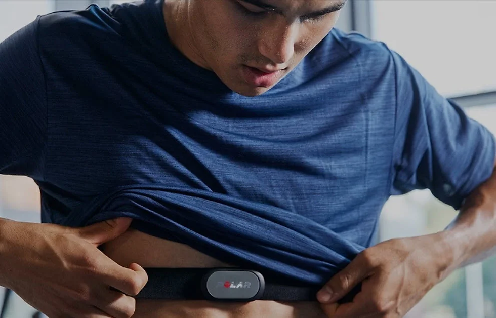
For training with smartphone applications like Optimal HRV, place an ECG sensor on your client's torso to avoid movement artifacts from forearm contraction.

The Polar H10 represents the gold standard for ambulatory ECG monitoring. Torso placement significantly reduces electromyographic (EMG) interference that commonly occurs with forearm sensors during muscle contraction exercises. This positioning ensures cleaner heart rate variability signals throughout your SPC training sessions.
SPC Training Using Data Acquisition Systems
Thought Technology Ltd.'s ProComp Infiniti system offers exceptional value in terms of accuracy, flexibility, and user-friendly software, like Dr. Inna Khazan's Mindfulness Suite.


Choose an ECG sensor, like Thought Technology Ltd.'s EKG-Flex/Pro. Use a chest or Erik Peper's lower torso placement, shown respectively. The lower torso placement preserves client modesty and reduces EMG contamination of the ECG signal.


Position
Clients should recline with their ankles crossed and feet supported by a footrest or separate chair. Although the original Vaschillo protocol only contracted wrists and ankles with legs uncrossed, we have observed greater respiratory sinus arrhythmia (RSA) using wrist, core, and crossed-ankle contraction. RSA represents breathing-mediated changes in peak-to-trough heart rate differences. The Truman Center for Applied Psychophysiology uses a Colamy office chair.

Training Protocol
Muscle Groups
Instruct your clients to gently simultaneously contract their wrists, core, and ankles.

Borrowing from Dr. Erik Peper's approach, encourage effortless contraction to ensure a smooth rhythm and minimize fatigue.

Your clients should use about 25% of their maximum effort. They should feel as if their wrists-core-ankles are contracting themselves, rather than forcing the movement.

This gentle approach promotes sustainable practice and prevents the activation of stress responses like vagal withdrawal that could counteract the training benefits.

They should hold each contraction for 3 seconds.

Below is an Optimal HRV breathing pacer we have repurposed for SPC. Clients should start 1.5 seconds before each cycle's peak. End 1.5 seconds after each cycle's peak.

This precise timing ensures optimal synchronization with the cardiovascular system's natural rhythms, maximizing the resonance effects that enhance heart rate variability.
Slow-Paced Contraction With or Without Slow-Paced Breathing
You can combine slow-paced contraction (SPC) with slow-paced breathing (SPB) or use it alone to increase HRV. When combining these techniques, limit contraction to the wrists and ankles to permit the abdomen to expand and the diaphragm to descend.
When Should You Combine SPC with SPB?
Dr. Inna Khazan combines SPC with SPB when her clients struggle to learn SPB. SPC can serve as a powerful pacing cue for their breathing rhythm, and model of the effortlessness we encourage.

Matt Bennett, Optimal HRV co-founder, recommends it to increase HRV for clients who have mastered SPB. He finds that their HR Max - HR Min and low-frequency values are greater when they combine SPC with SPB. They can see improved results after training trials using the Optimal HRV application

Several clinicians who treat Postural Orthostatic Tachycardia Syndrome (POTS), which is characterized by an excessive increase in heart rate upon standing, report that they have minimal HRV and require the combined approach to achieve clinical gains.
Data from the Truman Center for Applied Psychophysiology
The data were obtained from a healthy Truman undergraduate who had mastered slow-paced contraction and slow-paced breathing.
Time-Domain Measurements
The first Kubios table shows 6-cpm SPC. RMSSD was 55 ms.

The second table shows 6-cpm SPC with 6-bpm SPB. RMSSD was 88 ms.

Frequency-Domain Measurements
The first table shows 6-cpm SPC. Low-frequency power (ms2/Hz) using the FFT method was 5046. The participant breathed normally at 7.8 bpm.

The second table shows 6-cpm SPC with 6-bpm SPB. Low-frequency power (ms2/Hz) using the FFT method was 12277. The participant breathed at the target of 6 bpm.

Pilot Summary
These data suggest that two oscillators, 6-cpm SPC and 6-bpm SPB, can synergistically increase RSA and HRV time- and frequency-domain measurements. Confirmation awaits a planned Truman Center randomized controlled trial.
When Should You Use SPC Alone?
Prioritize slow-paced contraction (SPC) when slow-paced breathing (SPB) is not advisable due to dysfunctional breathing patterns or medical contraindications that make respiratory interventions unsafe or ineffective. SPC offers a means of engaging the autonomic nervous system through rhythmic, low-intensity skeletal muscle contractions that mimic the oscillatory effects of breathing on heart rate variability (HRV) and vagal modulation without directly altering respiration. This approach supports parasympathetic activation and enhances baroreflex engagement while maintaining safety for individuals with conditions such as chronic obstructive pulmonary disease (COPD), panic-prone respiratory sensitivity, or unstable cardiovascular responses. In such cases, SPC serves as a functional analog to SPB, providing a gentle, body-based entrainment method that stabilizes autonomic tone without invoking potentially disruptive ventilatory dynamics.
Dysfunctional Breathing
Slow-paced breathing can be challenging for clients who breathe dysfunctionally, particularly those who exhibit overbreathing or hyperventilation patterns. These individuals may struggle with the precise respiratory control required for effective SPB training.

SPC provides an alternative pathway to cardiovascular resonance that bypasses problematic breathing patterns, allowing these clients to access the benefits of HRV biofeedback training without respiratory-based interventions.
Medical Conditions
Elevated Respiratory Rates in Clinical Populations
Disorders that affect respiration may elevate breathing rates to 18-28 breaths per minute, according to research by Fried (1987) and Fried and Grimaldi (1993). These elevated rates make it extremely difficult for clients to slow their breathing to the 4.5-6.5 bpm range required for effective SPB training.
Conditions such as anxiety disorders and panic disorder can chronically elevate respiratory rates, making SPC a more accessible alternative intervention approach.

Pain-Related Respiratory Changes
Sustained pain increased the respiration rate from 13.2 to 17.7 bpm in research conducted by Kato, Kowalski, and Stohler (2001). Patients experiencing chronic pain may be unable to slow their breathing to the 4.5-6.5 bpm range necessary for effective SPB training.
Pain-related respiratory changes reflect the body's natural stress response and autonomic nervous system activation, making breathing-based interventions more challenging to implement effectively in these populations.

Metabolic Acidosis
SPB may be medically contraindicated when altered breathing patterns could be hazardous for clients suffering from diabetes (Kitabchi et al., 2009) or kidney disease (Kim, 2021) that produce metabolic acidosis—excess acid in the body fluid.
In these conditions, the body relies on specific respiratory patterns to maintain acid-base balance, making interventions that alter breathing potentially dangerous to the client's physiological homeostasis.

Respiratory Acidosis
Common respiratory acidosis causes include chronic obstructive pulmonary disease (COPD), asthma, pneumonia, and neuromuscular disorders that affect breathing muscles.
These conditions impair the lungs' ability to eliminate carbon dioxide effectively, leading to respiratory acidosis. Interventions that further alter breathing patterns could exacerbate these underlying physiological imbalances.

Patients may breathe rapidly to protect acid-base balance in medical disorders that cause a decrease in blood pH, leading to acidosis. Rapid breathing helps to expel carbon dioxide (CO2) from the body, which in turn can increase the pH and counteract the acidosis.

This compensatory hyperventilation represents a crucial physiological adaptation that maintains homeostasis. Interventions that interfere with this compensatory mechanism could compromise the patient's health and safety. SPC provides a safe alternative that can improve heart rate variability and autonomic balance without disrupting these essential respiratory compensatory mechanisms.
Key Takeaways
SPC is a core HRV exercise pioneered by Evgeny Vaschillo’s group; it can be paired with SPB or used instead when SPB is contraindicated (e.g., metabolic/kidney conditions).
Teach with a chest sensor (e.g., Polar H10) when using a smartphone app like Optimal HRV or with a three-lead ECG sensor lower-torso placement when using a data acquisition system like the ProComp Infiniti.
Position clients reclined, ankles crossed and supported; cue gentle, effortless wrist-core-ankle contractions held ~3 s. Encourage 25% effort to avoid vagal withdrawal.
Time each contraction to the pacer peak (start ~1.5 s before, end ~1.5 s after) to synchronize with cardiovascular rhythms and amplify resonance effects.
Combine SPC+SPB when appropriate and prioritize SPC alone for dysfunctional breathing or when slowing respiration would be unsafe.

Appreciation
The Truman Center for Applied Psychophysiology research staff made this post possible. A special thanks to my amazing Lab Managers, Isaac Compton and Emma Suchsland, who teach and supervise this dedicated team of 33 undergraduates. Isaac Compton modeled our wrists-core-ankles SPC technique.

Glossary
baroreflex: baroreceptor reflex that provides negative feedback control of BP. Elevated BP activates the baroreflex to lower BP, and low BP suppresses the baroreflex to raise BP.
effortless contraction: contraction force around 25% of maximum effort, analogous to Erik Peper's concept of effortless breathing.
HR Max – HR Min: an HRV index that calculates the average difference between the highest and lowest HRs during each respiratory cycle.
low-frequency (LF) band: a HRV frequency range of 0.04-0.15 Hz that may represent the influence of PNS and baroreflex activity when breathing or contracting muscles between 4.5-6.5 times a minute.
metabolic acidosis: a condition in which excess acid accumulates or bicarbonate is lost from body fluids, causing blood pH to fall below normal due to non-respiratory causes such as kidney dysfunction or diabetes.
respiratory acidosis: a condition in which the lungs fail to eliminate sufficient carbon dioxide (CO₂), leading to its buildup in the blood and a corresponding decrease in pH from impaired ventilation or gas exchange.
respiratory sinus arrhythmia (RSA): the respiration-driven heart rhythm that contributes to the high frequency (HF) component of heart rate variability. Inhalation inhibits vagal nerve slowing of the heart (increasing HR), while exhalation restores vagal slowing (decreasing HR).
RMSSD: the square root of the mean squared difference of adjacent NN intervals in milliseconds.
slow-paced breathing (SPB): breathing in the adult 4.5-6.5 breaths per minute range to stimulate cardiovascular resonance and increase heart rate variability.
slow-paced contraction (SPC): wrist-ankle or wrist-core-ankle contraction in the adult 4.5-6.5 contractions per minute range as an alternative to breathing-based HRV training.
vagal withdrawal: the reduction or inhibition of parasympathetic (vagal) influence on the heart, typically during stress, exercise, or cognitive demand. When the vagal input decreases—i.e., withdrawal occurs—heart rate increases, reflecting a shift from parasympathetic to sympathetic dominance.
References Fried, R. (1987). The hyperventilation syndrome: Research and clinical treatment. John Hopkins University Press.
Fried, R., & Grimaldi, J. (1993). The psychology and physiology of breathing. Springer. Goessl, V. C., Curtiss, J. E., & Hofmann, S. G. (2017). The effect of heart rate variability biofeedback training on stress and anxiety: A meta-analysis. Psychological medicine, 47(15), 2578–2586. https://doi.org/10.1017/S0033291717001003 Hansen, A. L., Johnsen, B. H., & Thayer, J. F. (2009). Relationship between heart rate variability and cognitive function during threat of shock. Anxiety, Stress, and Coping, 22(1), 77–89. https://doi.org/10.1080/10615800802272251 Kato, Y., Kowalski, C. J., & Stohler, C. S. (2001). Habituation of the early pain-specific respiratory response in sustained pain. Pain, 91(1-2), 57–63. https://doi.org/10.1016/s0304-3959(00)00419-x Kim, H. J. (2021). Metabolic acidosis in chronic kidney disease: Pathogenesis, clinical consequences, and treatment. Electrolyte & Blood Pressure: E & BP, 19(2), 29–37. https://doi.org/10.5049/EBP.2021.19.2.29
Kitabchi, A. E., Umpierrez, G. E., Miles, J. M., & Fisher, J. N. (2009). Hyperglycemic crises in adult patients with diabetes. Diabetes Care, 32(7), 1335–1343. https://doi.org/10.2337/dc09-9032 Lehrer, P. (2022). My life in HRV biofeedback research. Applied Psychophysiology and Biofeedback, 1-10. https://doi.org/10.1007/s10484-022-09535-5 Lehrer, P., Kaur, K., Sharma, A., Shah, K., Huseby, R., Bhavsar, J., Sgobba, P., & Zhang, Y. (2020). Heart rate variability biofeedback improves emotional and physical health and performance: A systematic review and meta-analysis. Applied Psychophysiology and Biofeedback, 45, 109-129. https://doi.org/10.1007/s10484-020-09466-z Meehan, Z. M., & Shaffer, F. (2023). Adding core muscle contraction to wrist-ankle rhythmical skeletal muscle tension increases respiratory sinus arrhythmia and low-frequency power. Applied Psychophysiology and Biofeedback, 48(1), 127–134. https://doi.org/10.1007/s10484-022-09568-w Shaffer, F., & Ginsberg, J. P. (2017). An overview of heart rate variability metrics and norms. Frontiers in Public Health. https://doi.org/10.3389/fpubh.2017.00258 Shaffer, F., Moss, D., & Meehan, Z. M. (2022). Rhythmic skeletal muscle tension increases heart rate variability at 1 and 6 contractions per minute. Appl Psychophysiol Biofeedback. https://doi.org/10.1007/s10484-022-09541-7 Vaschillo, E., Lehrer, P., Rishe, N., & Konstantinov, M. (2002). Heart rate variability biofeedback as a method for assessing baroreflex function: A preliminary study of resonance in the cardiovascular system. Applied Psychophysiology and Biofeedback, 27, 1-27. https://doi.org/10.1023/A:1014587304314
Vaschillo, E. G., Vaschillo, B., Pandina, R. J., & Bates, M. E. (2011). Resonances in the cardiovascular system caused by rhythmical muscle tension. Psychophysiology, 48, 927–936. https://doi.org/10.1111/j.1469-8986.2010.01156.x
About the Author
Fred Shaffer earned his PhD in Psychology from Oklahoma State University. He earned BCIA certifications in Biofeedback and HRV Biofeedback. Fred is an Allen Fellow and Professor of Psychology at Truman State University, where has has taught for 50 years. He is a Biological Psychologist who consults and lectures in heart rate variability biofeedback, Physiological Psychology, and Psychopharmacology. Fred helped to edit Evidence-Based Practice in Biofeedback and Neurofeedback (3rd and 4th eds.) and helped to maintain BCIA's certification programs.

Support Our Friends


Comments